Explore
 Validate
 Learn
Learn Western blot
 Immunocytochemistry
Antibody data
- Antibody Data
- Antigen structure
- References [1]
- Comments [0]
- Validations
- Immunocytochemistry [4]
- Immunohistochemistry [1]
Submit
Validation data
Reference
Comment
Report error
- Product number
- GTX22773 - Provider product page

- Provider
- GeneTex
- Proper citation
- GeneTex Cat#GTX22773, RRID:AB_384843
- Product name
- Prolactin Receptor antibody [T6]
- Antibody type
- Monoclonal
- Reactivity
- Human, Mouse, Rat
- Host
- Mouse
Submitted references Quercetin improves postpartum hypogalactia in milk-deficient mice via stimulating prolactin production in pituitary gland.
Lin M, Wang N, Yao B, Zhong Y, Lin Y, You T
Phytotherapy research : PTR 2018 Aug;32(8):1511-1520
Phytotherapy research : PTR 2018 Aug;32(8):1511-1520
No comments: Submit comment
Supportive validation
- Submitted by
- GeneTex (provider)
- Main image
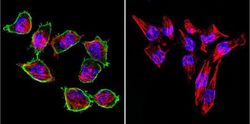
- Experimental details
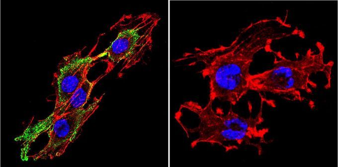
- Immunofluorescent analysis of Prolactin Receptor in H-4-II-E Cells. Cells were grown on chamber slides and fixed with formaldehyde prior to staining. Cells were probed without (control) or with a Prolactin Receptor monoclonal antibody at a dilution of 1:200 overnight at 4 C, washed with PBS and incubated with a DyLight-488 conjugated secondary antibody. Prolactin Receptor staining (green), F-Actin staining with Phalloidin (red) and nuclei with DAPI (blue) is shown. Images were taken at 60X magnification.

- Submitted by
- GeneTex (provider)
- Main image
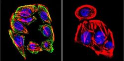
- Experimental details
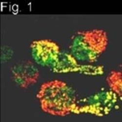
- Immunofluorescent analysis of Prolactin Receptor in SW480 Cells. Cells were grown on chamber slides and fixed with formaldehyde prior to staining. Cells were probed without (control) or with a Prolactin Receptor monoclonal antibody at a dilution of 1:200 overnight at 4 C, washed with PBS and incubated with a DyLight-488 conjugated secondary antibody. Prolactin Receptor staining (green), F-Actin staining with Phalloidin (red) and nuclei with DAPI (blue) is shown. Images were taken at 60X magnification.

- Submitted by
- GeneTex (provider)
- Main image
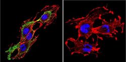
- Experimental details
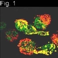
- Immunofluorescent analysis of Prolactin Receptor in C6 cells. Cells were grown on slides and fixed with formaldehyde prior to staining. Cells were probed without (control) or with Prolactin Receptor antibody [T6] at a dilution of 1:200 overnight at 4 C, washed with PBS and incubated with a proper secondary antibody. Prolactin Receptor staining (green), F-Actin staining with Phalloidin (red) and nuclei with DAPI (blue) is shown. Images were taken at 60X magnification.
- Submitted by
- GeneTex (provider)
- Main image
- Experimental details
- Immunofluorescent analysis of Prolactin Receptor in NB2 cells.
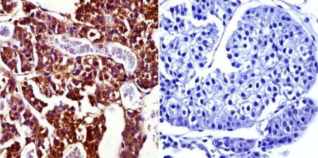
Supportive validation
- Submitted by
- GeneTex (provider)
- Main image
- Experimental details
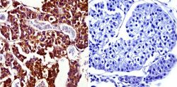
- Immunohistochemistry was performed on normal biopsies of deparaffinized Rat pituitary gland tissue. To expose target proteins, heat induced antigen retrieval was performed using 10mM sodium citrate (pH6.0) buffer, microwaved for 8-15 minutes. Following antigen retrieval tissues were blocked in 3% BSA-PBS for 30 minutes at room temperature and probed with a Prolactin Receptor monoclonal antibody at a dilution of 1:50 or without primary antibody (negative control) overnight at 4¢XC in a humidified chamber. Tissues were washed with PBST and endogenous peroxidase activity was quenched with a peroxidase suppressor. Detection was performed using a biotin-conjugated secondary antibody and SA-HRP, followed by colorimetric detection using DAB. Tissues were counterstained with hematoxylin and prepped for mounting.