Explore
 Validate
 Learn
Learn Western blot
Antibody data
- Antibody Data
- Antigen structure
- References [1]
- Comments [0]
- Validations
- Western blot [5]
- Immunocytochemistry [1]
- Immunohistochemistry [2]
Submit
Validation data
Reference
Comment
Report error
- Product number
- GTX106235 - Provider product page

- Provider
- GeneTex
- Proper citation
- GeneTex Cat#GTX106235, RRID:AB_2036504
- Product name
- CD2AP antibody
- Antibody type
- Polyclonal
- Reactivity
- Human, Mouse, Rat
- Host
- Rabbit
Submitted references CD2-Associated Protein Contributes to Hepatitis C, Virus Propagation and Steatosis by Disrupting Insulin Signaling.
Zhang H, Zhang C, Tang H, Gao S, Sun F, Yang Y, Zhou W, Hu Y, Ke C, Wu Y, Ding Z, Guo L, Pei R, Chen X, Sy MS, Zhang B, Li C
Hepatology (Baltimore, Md.) 2018 Nov;68(5):1710-1725
Hepatology (Baltimore, Md.) 2018 Nov;68(5):1710-1725
No comments: Submit comment
Supportive validation
- Submitted by
- GeneTex (provider)
- Main image
- Experimental details
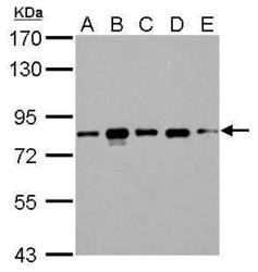
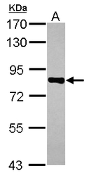
- CD2AP antibody detects CD2AP protein by western blot analysis.A. 30 ?g 293T whole cell lysate/extract B. 30 ?g A431 whole cell lysate/extract C. 30 ?g HeLa whole cell lysate/extract D. 30 ?g HepG2 whole cell lysate/extract E. 30 ?g A375 whole cell lysate/extract 7.5% SDS-PAGECD2AP antibody (GTX106235) dilution: 1:1000 The HRP-conjugated anti-rabbit IgG antibody (GTX213110-01) was used to detect the primary antibody.
- Submitted by
- GeneTex (provider)
- Main image
- Experimental details
- CD2AP antibody detects CD2AP protein by western blot analysis.A. 30 ?g Rat2 whole cell lysate/extract7.5% SDS-PAGECD2AP antibody (GTX106235) dilution: 1:1000 The HRP-conjugated anti-rabbit IgG antibody (GTX213110-01) was used to detect the primary antibody.
- Submitted by
- GeneTex (provider)
- Main image
- Experimental details
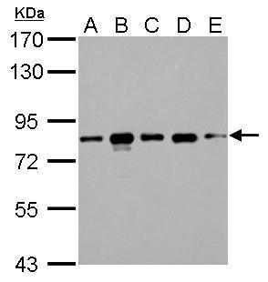
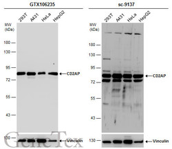
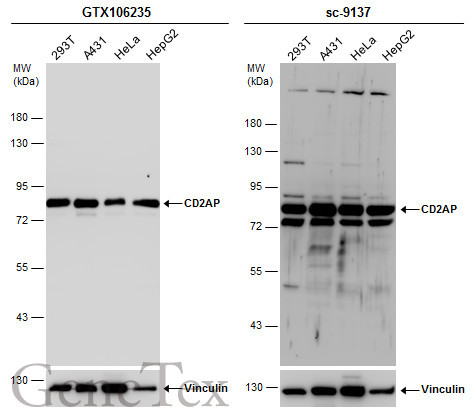
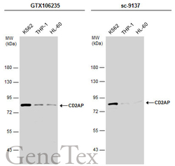
- Various whole cell extracts (30 ?g) were separated by 7.5% SDS-PAGE, and the membranes were blotted with CD2AP antibody (GTX106235) diluted at 1:1000 and competitor's antibody (sc-9137) diluted at 1:200. The HRP-conjugated anti-rabbit IgG antibody (GTX213110-01) was used to detect the primary antibody.*The competitor is not affiliated with GeneTex and does not endorse this product.
- Submitted by
- GeneTex (provider)
- Main image
- Experimental details
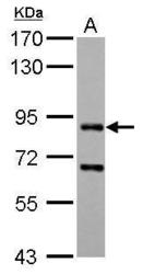
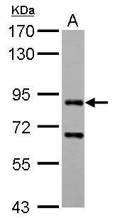
- Sample (30 ug of whole cell lysate) A: A431 7.5% SDS PAGE GTX106235 diluted at 1:1000

- Submitted by
- GeneTex (provider)
- Main image
- Experimental details
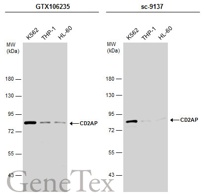
- Various whole cell extracts (30 ?g) were separated by 7.5% SDS-PAGE, and the membranes were blotted with CD2AP antibody (GTX106235) diluted at 1:1000 and competitor's antibody (sc-9137) diluted at 1:200. The HRP-conjugated anti-rabbit IgG antibody (GTX213110-01) was used to detect the primary antibody.
Supportive validation
- Submitted by
- GeneTex (provider)
- Main image
- Experimental details
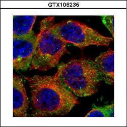
- Confocal immunofluorescence analysis (Olympus FV10i) of paraformaldehyde-fixed A431, using CD2AP(GTX106235) antibody (Green) at 1:500 dilution. Alpha-tubulin filaments were labeled with GTX11304 (Red) at 1:500.

Supportive validation
- Submitted by
- GeneTex (provider)
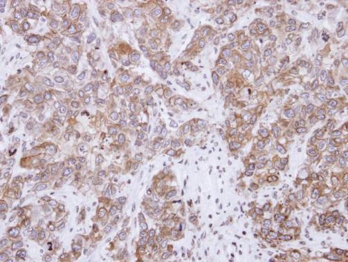
- Main image
- Experimental details
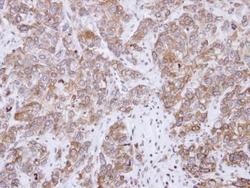
- Immunohistochemical analysis of paraffin-embedded FaDu xenograft, using CD2-associated protein(GTX106235) antibody at 1:500 dilution.
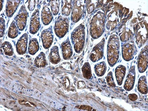
- Submitted by
- GeneTex (provider)
- Main image
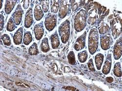
- Experimental details
- CD2AP antibody detects CD2AP protein at cytosol on mouse colon by immunohistochemical analysis. Sample: Paraffin-embedded mouse colon. CD2AP antibody (GTX106235) dilution: 1:500.