Explore
 Validate
 Learn
Learn Western blot
 Immunohistochemistry
Antibody data
- Antibody Data
- Antigen structure
- References [2]
- Comments [0]
- Validations
- Immunohistochemistry [6]
Submit
Validation data
Reference
Comment
Report error
- Product number
- HPA005835 - Provider product page

- Provider
- Atlas Antibodies
- Proper citation
- Atlas Antibodies Cat#HPA005835, RRID:AB_1847965
- Product name
- Anti-ECH1
- Antibody type
- Polyclonal
- Reactivity
- Human, Mouse, Rat
- Host
- Rabbit
- Conjugate
- Unconjugated
- Antigen sequence
EVDVGLAADVGTLQRLPKVIGNQSLVNELAFTARK
MMADEALGSGLVSRVFPDKEVMLDAALALAAEISS
KSPVAVQSTKVNLLYSRDHSVAESLNYVASWNMSM
LQTQDLVKSVQATTENKELKTVTF- Isotype
- IgG
- Vial size
- 100 µl
- Storage
- Store at +4°C for short term storage. Long time storage is recommended at -20°C.
Submitted references Systematic validation of antibody binding and protein subcellular localization using siRNA and confocal microscopy
Tissue profiling of the mammalian central nervous system using human antibody-based proteomics.
Stadler C, Hjelmare M, Neumann B, Jonasson K, Pepperkok R, Uhlén M, Lundberg E
Journal of Proteomics 2012 April;75(7):2236-2251
Journal of Proteomics 2012 April;75(7):2236-2251
Tissue profiling of the mammalian central nervous system using human antibody-based proteomics.
Mulder J, Björling E, Jonasson K, Wernérus H, Hober S, Hökfelt T, Uhlén M
Molecular & cellular proteomics : MCP 2009 Jul;8(7):1612-22
Molecular & cellular proteomics : MCP 2009 Jul;8(7):1612-22
No comments: Submit comment
Supportive validation
- Submitted by
- Atlas Antibodies (provider)
- Main image
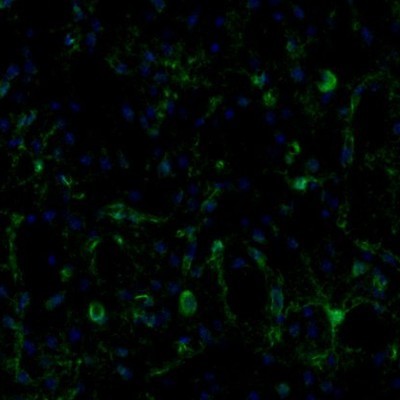
- Experimental details
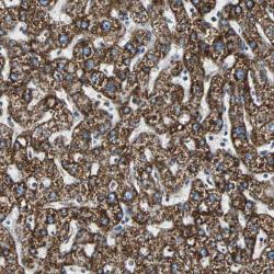
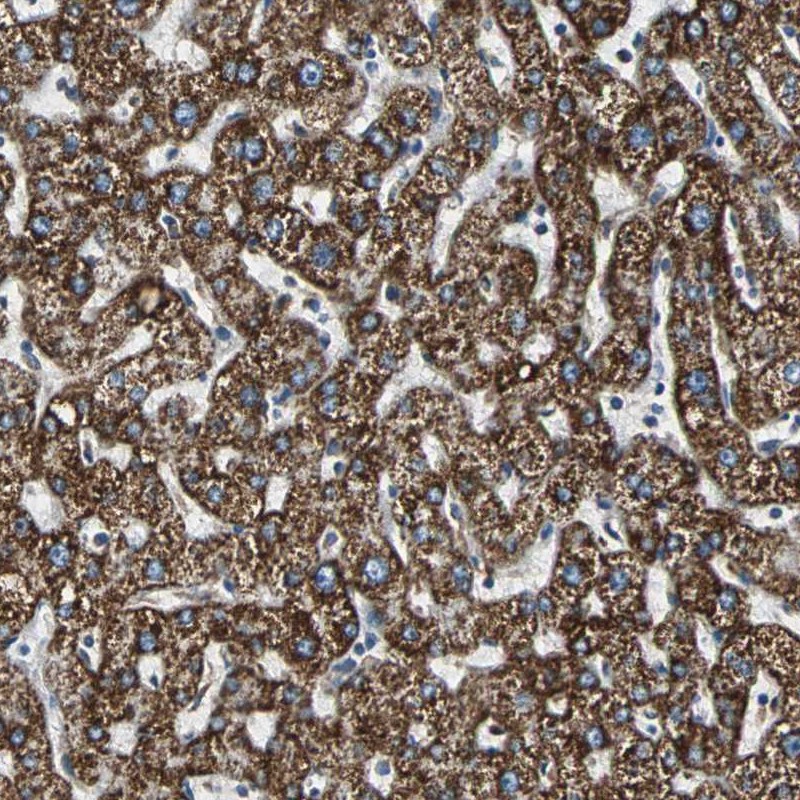
- Immunohistochemical staining of human liver shows strong cytoplasmic positivity in a granular pattern in hepatocytes.
- Sample type
- HUMAN
- Submitted by
- Atlas Antibodies (provider)
- Main image
- Experimental details
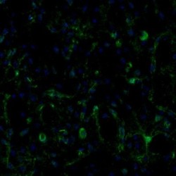
- Immunofluorescence staining of mouse olfactory bulb shows positivity in the mitral cell layer.
- Sample type
- MOUSE
- Submitted by
- Atlas Antibodies (provider)
- Main image
- Experimental details
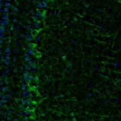
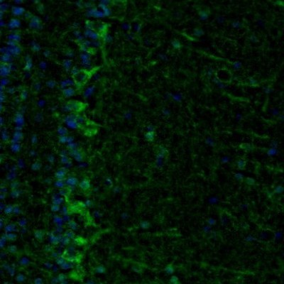
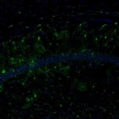
- Immunofluorescence staining of mouse caudate putamen shows positivity in neurons and blood vessels.
- Sample type
- MOUSE
- Submitted by
- Atlas Antibodies (provider)
- Main image
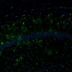
- Experimental details
- Immunofluorescence staining of mouse hippocampus shows immunoreactivity in the CA1 region.
- Sample type
- MOUSE
- Submitted by
- Atlas Antibodies (provider)
- Main image
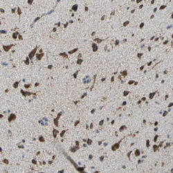
- Experimental details
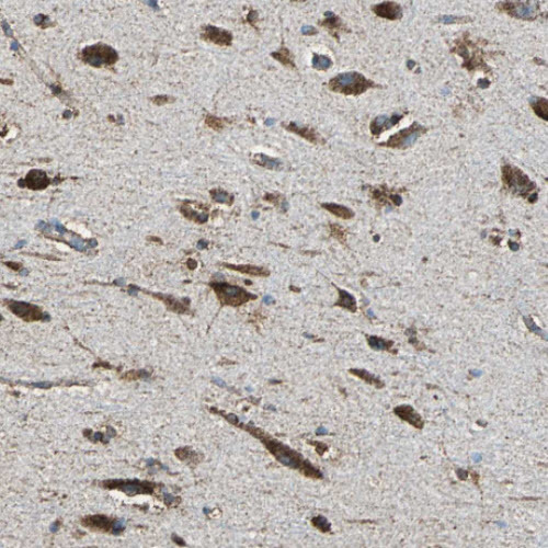
- Immunohistochemical staining of human cerebral cortex shows strong cytoplasmic immunoreactivity in neurons.
- Sample type
- HUMAN

- Submitted by
- Atlas Antibodies (provider)
- Main image

- Experimental details
- Immunohistochemical staining of human hippocampus shows strong cytoplasmic positivity in neuronal cell bodies.
- Sample type
- HUMAN