 Explore
 Validate
 Learn
LearnHPA001897
antibody from Atlas Antibodies
Targeting: ILF3
DRBP76, MPHOSPH4, MPP4, MPP4110, NF110, NF110b, NF90, NF90a, NF90c, NF90ctv, NFAR-1, NFAR-2, NFAR110, NFAR90, TCP110
 Western blot
 Immunocytochemistry
Antibody data
- Antibody Data
- Antigen structure
- References [2]
- Comments [0]
- Validations
- Immunocytochemistry [1]
- Immunohistochemistry [5]
Submit
Validation data
Reference
Comment
Report error
- Product number
- HPA001897 - Provider product page

- Provider
- Atlas Antibodies
- Proper citation
- Atlas Antibodies Cat#HPA001897, RRID:AB_1078703
- Product name
- Anti-ILF3
- Antibody type
- Polyclonal
- Reactivity
- Human
- Host
- Rabbit
- Conjugate
- Unconjugated
- Antigen sequence
VMAKHSSVYPTQEELEAVQNMVSHTERALKAVSDW
IDEQEKGSSEQAESDNMDVPPEDDSKEGAGEQKTE
HMTRTLRGVMRVGLVAKGLLLKGDLDLELVLLCKE
KPTTALLDKVADNLAIQLAAVTEDKYEILQSVDDA
AIVIK- Isotype
- IgG
- Vial size
- 100 µl
- Storage
- Store at +4°C for short term storage. Long time storage is recommended at -20°C.
Submitted references Sequestration by IFIT1 impairs translation of 2'O-unmethylated capped RNA.
Systematic validation of antibody binding and protein subcellular localization using siRNA and confocal microscopy
Habjan M, Hubel P, Lacerda L, Benda C, Holze C, Eberl CH, Mann A, Kindler E, Gil-Cruz C, Ziebuhr J, Thiel V, Pichlmair A
PLoS pathogens 2013;9(10):e1003663
PLoS pathogens 2013;9(10):e1003663
Systematic validation of antibody binding and protein subcellular localization using siRNA and confocal microscopy
Stadler C, Hjelmare M, Neumann B, Jonasson K, Pepperkok R, Uhlén M, Lundberg E
Journal of Proteomics 2012 April;75(7):2236-2251
Journal of Proteomics 2012 April;75(7):2236-2251
No comments: Submit comment
Supportive validation
- Submitted by
- Atlas Antibodies (provider)
- Main image
- Experimental details
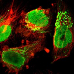
- Immunofluorescent staining of human cell line U-251 MG shows localization to nucleoplasm, nucleoli & mitochondria.
- Sample type
- HUMAN

Supportive validation
- Submitted by
- Atlas Antibodies (provider)
- Main image

- Experimental details
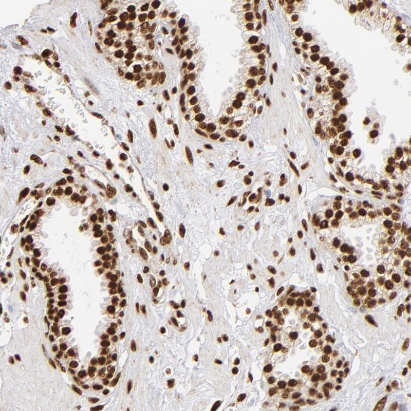
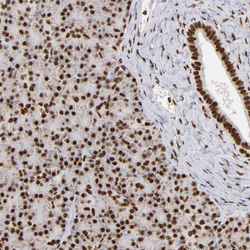
- Immunohistochemical staining of human prostate shows strong nuclear positivity in glandular cells.
- Submitted by
- Atlas Antibodies (provider)
- Main image
- Experimental details
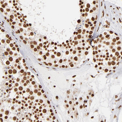
- Immunohistochemical staining of human lymph node shows strong nuclear positivity in cells in seminiferous ducts.
- Sample type
- HUMAN

- Submitted by
- Atlas Antibodies (provider)
- Main image
- Experimental details
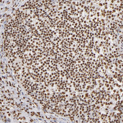
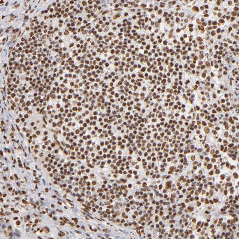
- Immunohistochemical staining of human lymph node shows strong nuclear positivity in germinal center cells.
- Sample type
- HUMAN
- Submitted by
- Atlas Antibodies (provider)
- Main image
- Experimental details
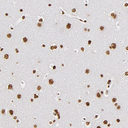
- Immunohistochemical staining of human cerebral cortex shows strong nuclear positivity in neurons.
- Sample type
- HUMAN
- Submitted by
- Atlas Antibodies (provider)
- Main image
- Experimental details
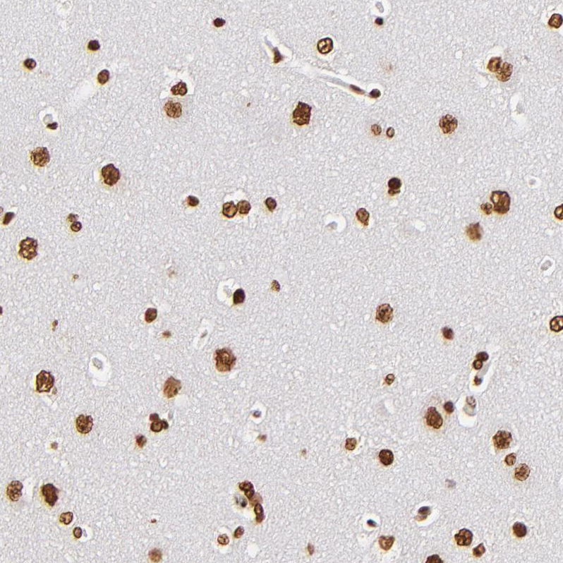
- Immunohistochemical staining of human pancreas shows strong nuclear positivity in exocrine glandular cells.
- Sample type
- HUMAN
