 Explore
 Validate
 Learn
Learn Western blot
 ELISA
ELISAAntibody data
- Antibody Data
- Antigen structure
- References [0]
- Comments [0]
- Validations
- Western blot [5]
- Immunocytochemistry [3]
- Immunohistochemistry [4]
- Flow cytometry [2]
Submit
Validation data
Reference
Comment
Report error
- Product number
- MA5-15899 - Provider product page

- Provider
- Invitrogen Antibodies
- Product name
- Prohibitin Monoclonal Antibody (5H7)
- Antibody type
- Monoclonal
- Antigen
- Purifed from natural sources
- Description
- MA5-15899 targets PHB in indirect ELISA, FACS, ICC, IHC, IF and WB applications and shows reactivity with Human, mouse, Non-human primate, and Rat samples. The MA5-15899 immunogen is purified recombinant fragment of human PHB expressed in E. Coli. . MA5-15899 detects PHB which has a predicted molecular weight of approximately 30kDa.
- Reactivity
- Human, Mouse, Rat
- Host
- Mouse
- Isotype
- IgG
- Antibody clone number
- 5H7
- Vial size
- 100 µL
- Concentration
- Conc. Not Determined
- Storage
- Store at 4°C short term. For long term storage, store at -20°C, avoiding freeze/thaw cycles.
No comments: Submit comment
Supportive validation
- Submitted by
- Invitrogen Antibodies (provider)
- Main image

- Experimental details
- Western blot analysis of PHB using a PHB monoclonal antibody (Product # MA5-15899) against a human PHB (AA: 68-259) recombinant protein.

- Submitted by
- Invitrogen Antibodies (provider)
- Main image

- Experimental details
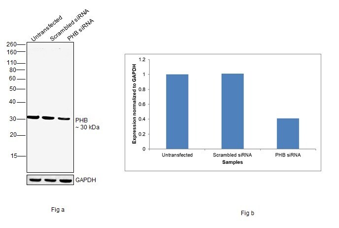
- Knockdown of Prohibitin was achieved by transfecting A-431 with Prohibitin specific siRNAs (Silencer® select Product # s10425, s10424). Western blot analysis (Fig. a) was performed using Whole cell extracts from the Prohibitin knockdown cells (lane 3), non-targeting scrambled siRNA transfected cells (lane 2) and untransfected cells (lane 1). The blot was probed with Prohibitin Monoclonal Antibody (5H7) (Product # MA5-15899, 1:1000 dilution) and Goat anti-Mouse IgG (H+L) Superclonal™ Recombinant Secondary Antibody, HRP (Product # A28177, 1:4000 dilution). Densitometric analysis of this western blot is shown in histogram (Fig. b). Decrease in signal upon siRNA mediated knock down confirms that antibody is specific to Prohibitin.
- Submitted by
- Invitrogen Antibodies (provider)
- Main image
- Experimental details
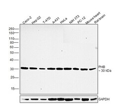
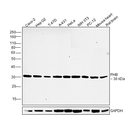
- Western blot was performed using Anti-Prohibitin Monoclonal Antibody (5H7)(Product # MA5-15899) and a 30 kDa band corresponding to Prohibitin was observed across cell lines and tissues tested. Whole cell extracts (30 µg lysate) of Caco-2 (Lane 1), Hep G2 (Lane 2), T-47D (Lane 3), A-431 (Lane 4), HeLa (Lane 5), NIH/3T3 (Lane 6), PC-12 (Lane 7) and tissue extracts (30 µg lysate) of Mouse Heart (Lane 8) and Rat Brain (Lane 9) were electrophoresed using NuPAGE™ 12% Bis-Tris Protein Gel (Product # NP0341BOX). Resolved proteins were then transferred onto a Nitrocellulose membrane (Product # IB23001) by iBlot® 2 Dry Blotting System (Product # IB21001). The blot was probed with the primary antibody (1:1000 dilution) and detected by chemiluminescence with Goat anti-Mouse IgG (H+L) Superclonal™ Recombinant Secondary Antibody, HRP (Product # A28177,1:4000 dilution) using the iBright FL 1000 (Product # A32752). Chemiluminescent detection was performed using Novex® ECL Chemiluminescent Substrate Reagent Kit (Product # WP20005).
- Submitted by
- Invitrogen Antibodies (provider)
- Main image

- Experimental details
- Western blot analysis of PHB using a PHB monoclonal antibody (Product # MA5-15899) against a human PHB (AA: 68-259) recombinant protein.

- Submitted by
- Invitrogen Antibodies (provider)
- Main image
- Experimental details
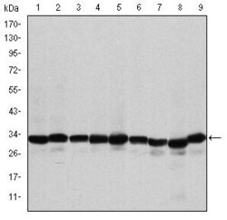
- Western blot analysis of PHB using PHB monoclonal antibody (Product # MA5-15899) in A431 (1), MCF-7 (2), Jurkat (3), HeLa (4), HepG2 (5), A549 (6), NIH/3T3 (7), COS-7 (8) and PC-12 (9) cell lysate.

Supportive validation
- Submitted by
- Invitrogen Antibodies (provider)
- Main image

- Experimental details
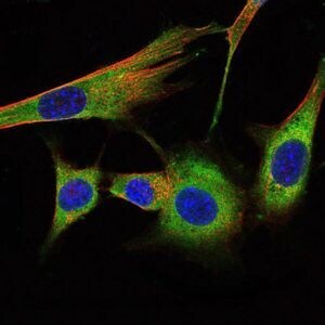
- Immunofluorescence analysis of NIH/3T3 cells using PHB monoclonal antibody (Product # MA5-15899) (Green). Blue: DRAQ5 fluorescent DNA dye. Red: actin filaments have been labeled with phalloidin.

- Submitted by
- Invitrogen Antibodies (provider)
- Main image
- Experimental details
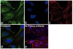
- Immunofluorescence analysis of Prohibitin was performed using 70% confluent log phase HeLa cells. The cells were fixed with 4% paraformaldehyde for 10 minutes, permeabilized with 0.1% Triton™ X-100 for 15 minutes, and blocked with 2% BSA for 45 minutes at room temperature. The cells were labeled with Prohibitin Monoclonal Antibody (5H7) (Product # MA5-15899) at 1:500 dilution in 0.1% BSA, incubated at 4 degree celsius overnight and then labeled with Goat anti-Mouse IgG (H+L) Highly Cross-Adsorbed Secondary Antibody, Alexa Fluor Plus 488 (Product # A32723), (1:2000 dilution), for 45 minutes at room temperature (Panel a: Green). Nuclei (Panel b:Blue) were stained with ProLong™ Diamond Antifade Mountant with DAPI (Product # P36962). F-actin (Panel c: Red) was stained with Rhodamine Phalloidin (Product # R415, 1:300 dilution). Panel d represents the merged image showing cytoplasm and nucleus localization. Panel e represents control cells with no primary antibody to assess background. The images were captured at 60X magnification.

- Submitted by
- Invitrogen Antibodies (provider)
- Main image

- Experimental details
- Immunofluorescence analysis of NIH/3T3 cells using PHB monoclonal antibody (Product # MA5-15899) (Green). Blue: DRAQ5 fluorescent DNA dye. Red: actin filaments have been labeled with phalloidin.
Supportive validation
- Submitted by
- Invitrogen Antibodies (provider)
- Main image

- Experimental details
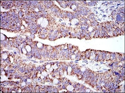
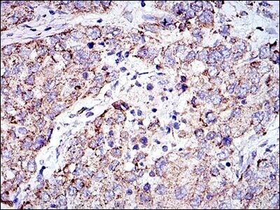
- Immunohistochemical analysis of paraffin-embedded rectum cancer tissues using PHB monoclonal antibody (Product # MA5-15899) followed with DAB staining.
- Submitted by
- Invitrogen Antibodies (provider)
- Main image

- Experimental details
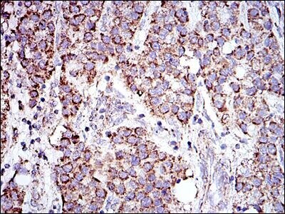
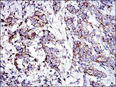
- Immunohistochemical analysis of paraffin-embedded liver cancer tissues using PHB monoclonal antibody (Product # MA5-15899) followed with DAB staining.
- Submitted by
- Invitrogen Antibodies (provider)
- Main image

- Experimental details
- Immunohistochemical analysis of paraffin-embedded lung cancer tissues using PHB monoclonal antibody (Product # MA5-15899) followed with DAB staining.
- Submitted by
- Invitrogen Antibodies (provider)
- Main image

- Experimental details
- Immunohistochemical analysis of paraffin-embedded stomach cancer tissues using PHB monoclonal antibody (Product # MA5-15899) followed with DAB staining.
Supportive validation
- Submitted by
- Invitrogen Antibodies (provider)
- Main image

- Experimental details
- Flow cytometric analysis of MCF-7 cells using PHB monoclonal antibody (Product # MA5-15899) (blue) and negative control (red).

- Submitted by
- Invitrogen Antibodies (provider)
- Main image

- Experimental details
- Flow cytometric analysis of MCF-7 cells using PHB monoclonal antibody (Product # MA5-15899) (blue) and negative control (red).
