Explore
 Validate
 Learn
Learn Western blot
 Immunohistochemistry
Antibody data
- Antibody Data
- Antigen structure
- References [0]
- Comments [0]
- Validations
- Western blot [1]
- Immunohistochemistry [8]
Submit
Validation data
Reference
Comment
Report error
- Product number
- HPA063181 - Provider product page

- Provider
- Atlas Antibodies
- Proper citation
- Atlas Antibodies Cat#HPA063181, RRID:AB_2684955
- Product name
- Anti-GZMK
- Antibody type
- Polyclonal
- Reactivity
- Human
- Host
- Rabbit
- Conjugate
- Unconjugated
- Antigen sequence
KCKVTGWGATDPDSLRPSDTLREVTVTVLSRKLCN
SQSYYNGDPFITKDMVCAGDAKGQKDS- Isotype
- IgG
- Vial size
- 100 µl
- Storage
- Store at +4°C for short term storage. Long time storage is recommended at -20°C.
No comments: Submit comment
Supportive validation
- Submitted by
- Atlas Antibodies (provider)
- Main image
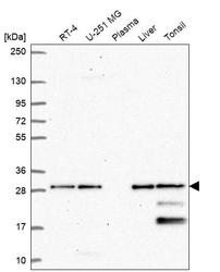
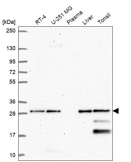
- Experimental details
- Western blot analysis in human cell line RT-4, human cell line U-251 MG, human plasma, human liver tissue and human tonsil tissue.
Enhanced validation
Supportive validation
- Submitted by
- Atlas Antibodies (provider)
- Enhanced method
- Orthogonal validation
- Main image
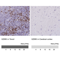
- Experimental details
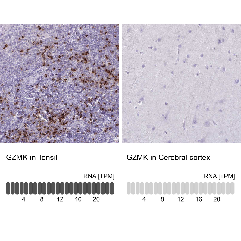
- Immunohistochemistry analysis in human tonsil and cerebral cortex tissues using HPA063181 antibody. Corresponding GZMK RNA-seq data are presented for the same tissues.
- Sample type
- HUMAN
Supportive validation
- Submitted by
- Atlas Antibodies (provider)
- Main image

- Experimental details
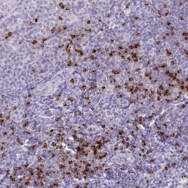
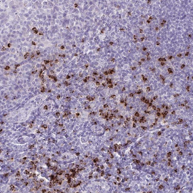
- Immunohistochemical staining of human tonsil shows high expression.
- Sample type
- HUMAN
- Submitted by
- Atlas Antibodies (provider)
- Main image
- Experimental details
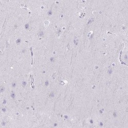
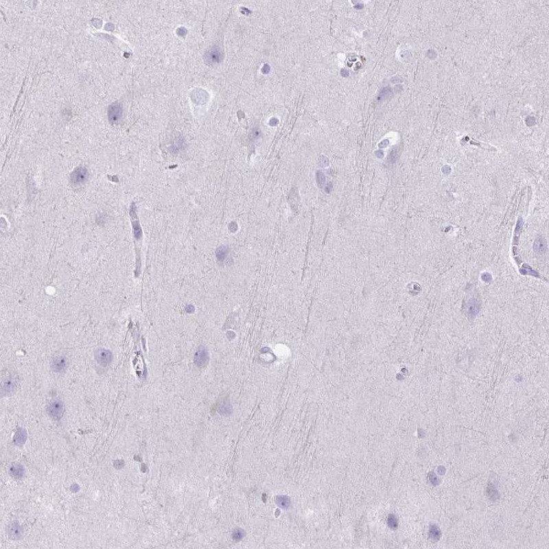
- Immunohistochemical staining of human cerebral cortex shows low expression as expected.
- Sample type
- HUMAN
- Submitted by
- Atlas Antibodies (provider)
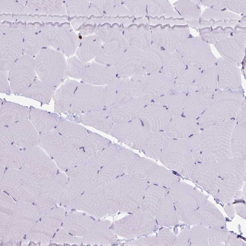
- Main image
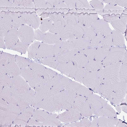
- Experimental details
- Immunohistochemical staining of human skeletal muscle shows no positivity in myocytes as expected.
- Sample type
- HUMAN
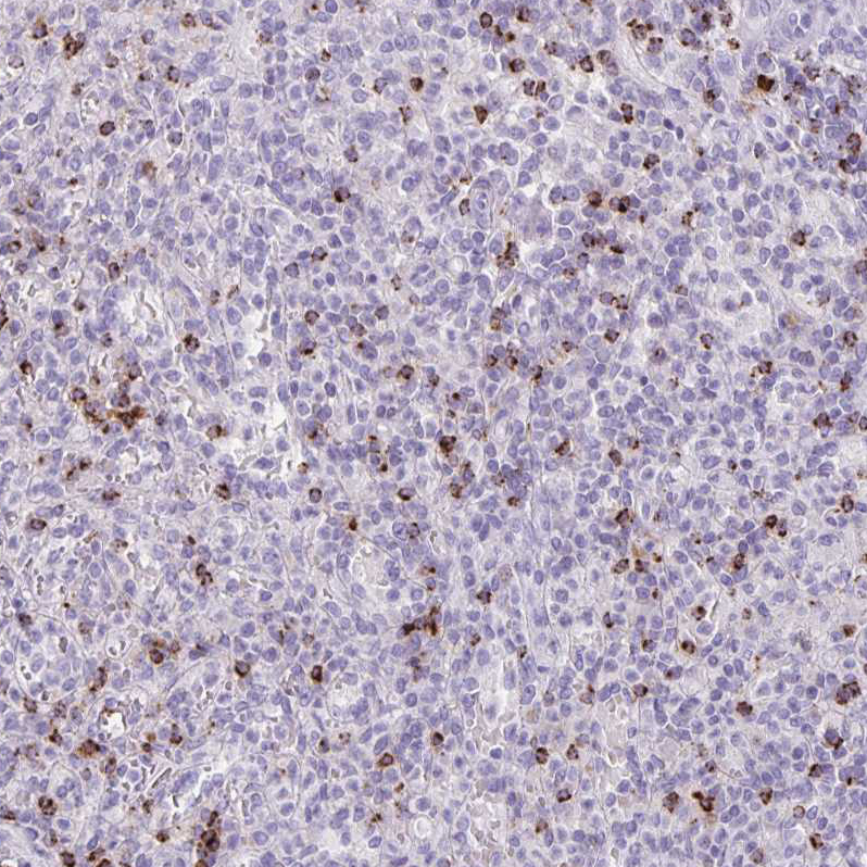
- Submitted by
- Atlas Antibodies (provider)
- Main image
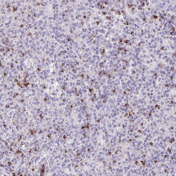
- Experimental details
- Immunohistochemical staining of human spleen shows strong granular cytoplasmic positivity in cells in red pulp.
- Sample type
- HUMAN
- Submitted by
- Atlas Antibodies (provider)
- Main image
- Experimental details
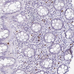
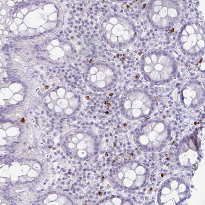
- Immunohistochemical staining of human rectum shows weak granular cytoplasmic positivity in leukocytes.
- Sample type
- HUMAN
- Submitted by
- Atlas Antibodies (provider)
- Main image
- Experimental details
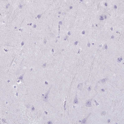
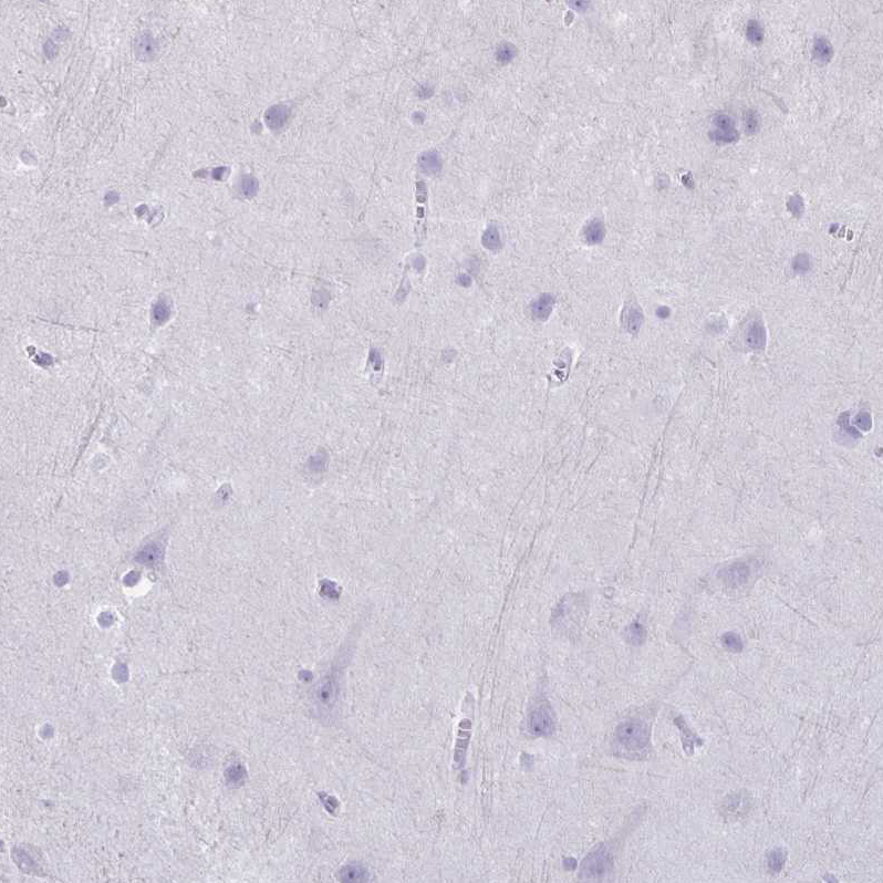
- Immunohistochemical staining of human cerebral cortex shows no positivity in neurons as expected.
- Sample type
- HUMAN
- Submitted by
- Atlas Antibodies (provider)
- Main image

- Experimental details
- Immunohistochemical staining of human tonsil shows strong granular cytoplasmic positivity in non-germinal center cells.
- Sample type
- HUMAN