Explore
 Validate
 Learn
Learn Western blot
Antibody data
- Antibody Data
- Antigen structure
- References [0]
- Comments [0]
- Validations
- Western blot [2]
- Immunohistochemistry [10]
- Other assay [1]
Submit
Validation data
Reference
Comment
Report error
- Product number
- UM800092 - Provider product page

- Provider
- OriGene
- Proper citation
- OriGene Cat#UM800092, RRID:AB_2629201
- Product name
- PROCR mouse monoclonal antibody,clone UMAB200
- Antibody type
- Monoclonal
- Description
- PROCR mouse monoclonal antibody,clone UMAB200
- Host
- Mouse
- Conjugate
- Unconjugated
- Epitope
- PROCR
- Isotype
- IgG
- Antibody clone number
- UMAB200
- Vial size
- 100 µl
- Concentration
- 1.00mg/ml
No comments: Submit comment
Supportive validation
- Submitted by
- OriGene (provider)
- Main image
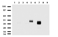
- Experimental details
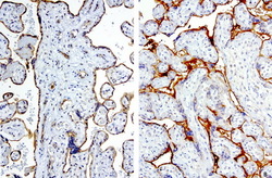
- Western blot of cell lysates (35ug) from 9 different cell lines (1: HepG2, 2: HeLa, 3: SV-T2, 4: A549. 5: COS7, 6: Jurkat, 7: MDCK, 8: PC-12, 9: MCF7).
- Validation comment
- WB

- Submitted by
- OriGene (provider)
- Main image
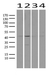
- Experimental details
- Western blot analysis of extracts (25ug) from 4 different cell lines by using anti-PROCR monoclonal antibody. (1:500) (1: HEK293; 2: Hela; 3: MCF7; 4: HepG2) Dilution: 1:500
- Validation comment
- WB

Supportive validation
- Submitted by
- OriGene (provider)
- Main image
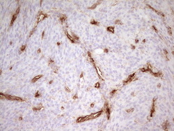
- Experimental details
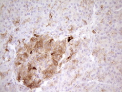
- Immunohistochemical staining of paraffin-embedded Human endometrium tissue using anti-PROCR mouse monoclonal antibody. (Heat-induced epitope retrieval by 1mM EDTA in 10mM Tris buffer (pH8.0) at 120C for 3 min, UM800092)(1:200)
- Validation comment
- IHC

- Submitted by
- OriGene (provider)
- Main image
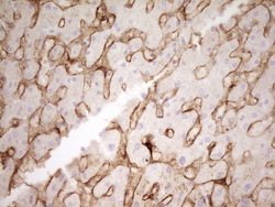
- Experimental details
- Immunohistochemical staining of paraffin-embedded Human pancreas tissue using anti-PROCR mouse monoclonal antibody. (Heat-induced epitope retrieval by 1mM EDTA in 10mM Tris buffer (pH8.0) at 120C for 3 min, UM800092)(1:200)
- Validation comment
- IHC

- Submitted by
- OriGene (provider)
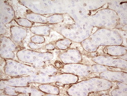
- Main image
- Experimental details
- Immunohistochemical staining of paraffin-embedded Human liver tissue using anti-PROCR mouse monoclonal antibody.(Heat-induced epitope retrieval by 1mM EDTA in 10mM Tris buffer (pH8.0) at 120C for 3 min, UM800092)(1:200)
- Validation comment
- IHC

- Submitted by
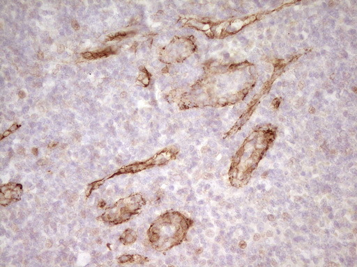
- OriGene (provider)
- Main image
- Experimental details
- Immunohistochemical staining of paraffin-embedded Carcinoma of Human liver tissue using anti-PROCR mouse monoclonal antibody.(Heat-induced epitope retrieval by 1mM EDTA in 10mM Tris buffer (pH8.0) at 120C for 3 min, UM800092)(1:200)
- Validation comment
- IHC

- Submitted by
- OriGene (provider)
- Main image

- Experimental details
- Immunohistochemical staining of paraffin-embedded Human tonsil using anti-PROCR mouse monoclonal antibody.(Heat-induced epitope retrieval by 1mM EDTA in 10mM Tris buffer (pH8.0) at 120C for 3 min, UM800092)(1:200)
- Validation comment
- IHC
- Submitted by
- OriGene (provider)
- Main image
- Experimental details
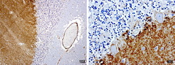
- Immunohistochemical staining of paraffin-embedded human brain using anti-PROCR clone UMAB200 mouse monoclonal antibody (UM800092) at 1:100 with Polink2 Broad HRP DAB detection kit; heat-induced epitope retrieval with GBI citrate pH6.0 HIER buffer using pressure chamber for 3 minutes at 110C. Very strong cytoplasmic and membraneous staining in the molecular layer.
- Validation comment
- IHC

- Submitted by
- OriGene (provider)
- Main image

- Experimental details
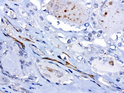
- Immunohistochemical staining of paraffin-embedded human thyroid cancer using anti-PROCR clone UMAB200 mouse monoclonal antibody (UM800092) at 1:100 with Polink2 Broad HRP DAB detection kit; heat-induced epitope retrieval with GBI citrate pH6.0 HIER buffer using pressure chamber for 3 minutes at 110C. Cytoplasmic staining is very strong in the vascular endothelia cells no staining in tumor cells.
- Validation comment
- IHC
- Submitted by
- OriGene (provider)
- Main image

- Experimental details
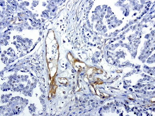
- Immunohistochemical staining of paraffin-embedded human ovarian cancer using anti-PROCR clone UMAB200 mouse monoclonal antibody (UM800092) at 1:100 with Polink2 Broad HRP DAB detection kit; heat-induced epitope retrieval with GBI citrate pH6.0 HIER buffer using pressure chamber for 3 minutes at 110C. Cytoplasmic staining is very strong in the vascular endothelia cells and weak nuclear staining in ovarian tumor cells.
- Validation comment
- IHC
- Submitted by
- OriGene (provider)
- Main image
- Experimental details
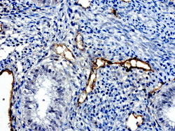
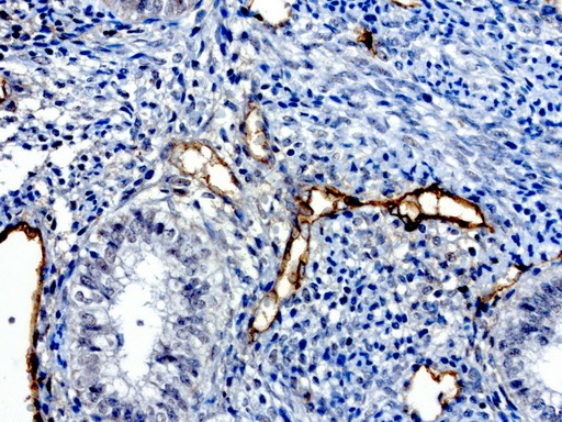
- Immunohistochemical staining of paraffin-embedded human normal adjacent endometrium using anti-PROCR clone UMAB200 mouse monoclonal antibody (UM800092) at 1:100 with Polink2 Broad HRP DAB detection kit; heat-induced epitope retrieval with GBI citrate pH6.0 HIER buffer using pressure chamber for 3 minutes at 110C. Cytoplasmic staining is very strong in the vascular endothelia cells and weak nuclear and cytoplasmic staining in in the glandular endometrial cells.
- Validation comment
- IHC
- Submitted by
- OriGene (provider)
- Main image
- Experimental details
- Immunohistochemical staining of 2 cases of paraffin-embedded human placenta using anti-PROCR clone UMAB200 mouse monoclonal antibody (UM800092) at 1:100 with Polink2 Broad HRP DAB detection kit; heat-induced epitope retrieval with GBI citrate pH6.0 HIER buffer using pressure chamber for 3 minutes at 110C. Very strong membraneous staining in the trophoblast cells. The endothelial cells show both cytoplasmic and membraneous staining.
- Validation comment
- IHC

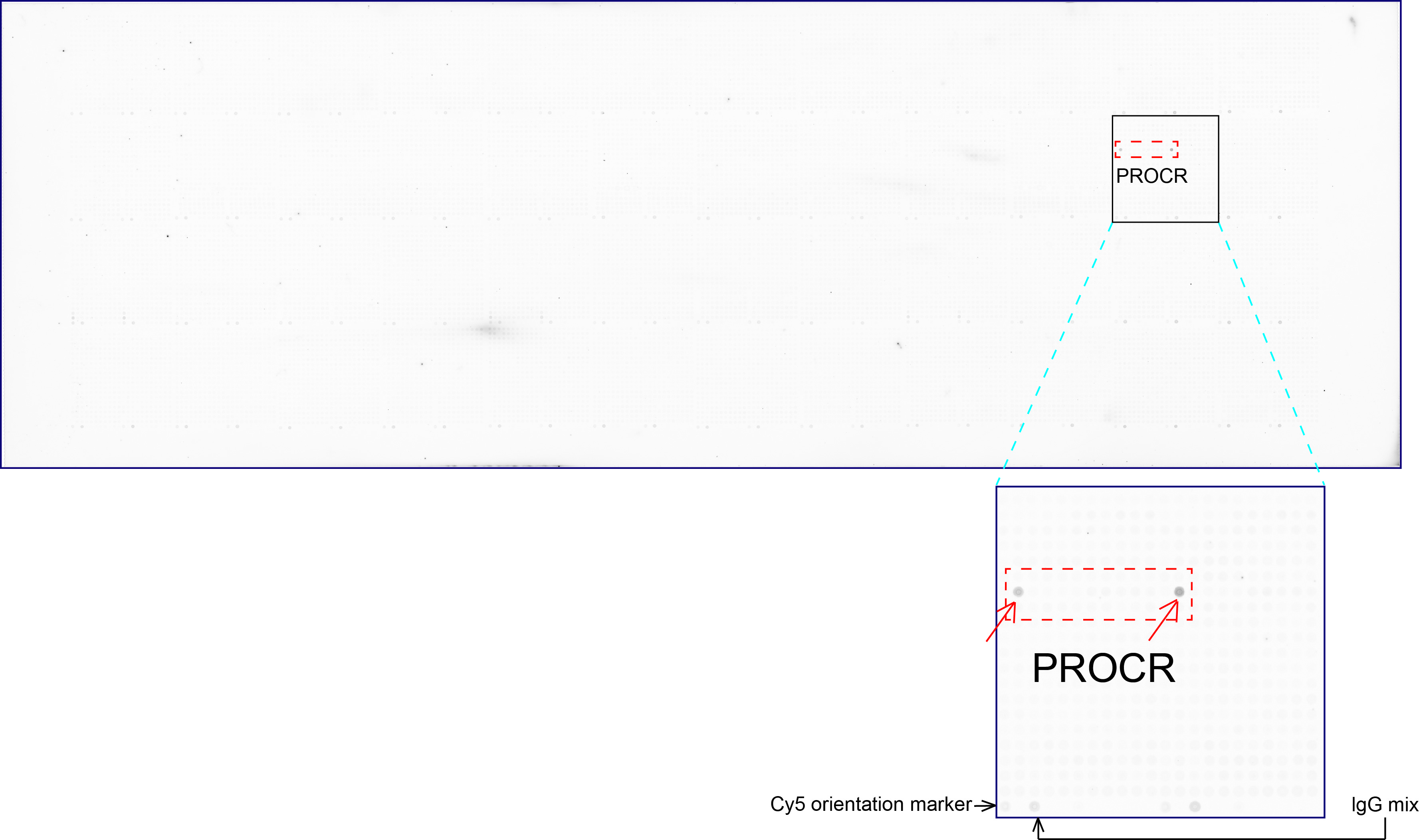
Supportive validation
- Submitted by
- OriGene (provider)
- Main image
- Experimental details
- OriGene overexpression protein microarray chip was immunostained with UltraMAB anti-PROCR mouse monoclonal antibody (UM800092). The positive reactive proteins are highlighted with two red arrows in the enlarged subarray. All the positive controls spotted in this subarray are also labeled for clarification.(1:100)
- Validation comment
- 10K-CHIP