Explore
 Validate
 Learn
Learn Western blot
 Immunocytochemistry
Antibody data
- Antibody Data
- Antigen structure
- References [0]
- Comments [0]
- Validations
- Western blot [2]
- Immunocytochemistry [2]
- Immunohistochemistry [2]
- Chromatin Immunoprecipitation [1]
Submit
Validation data
Reference
Comment
Report error
- Product number
- GTX54571 - Provider product page

- Provider
- GeneTex
- Product name
- PRDM14 antibody
- Antibody type
- Polyclonal
- Reactivity
- Human, Rat
- Host
- Rabbit
No comments: Submit comment
Enhanced validation
Supportive validation
- Submitted by
- GeneTex (provider)
- Enhanced method
- Genetic validation
- Main image

- Experimental details
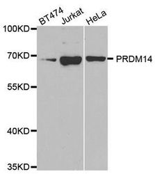
- The membrane was blotted with PRDM14 antibody (GTX54571) diluted by 1:1000.
- Protocol
- Protocol
- Knockdown/Genetic Approaches Application
- Western blot

Supportive validation
- Submitted by
- GeneTex (provider)
- Main image
- Experimental details
- WB analysis of extracts from various cells using PRDM14 antibody.
- Validation comment
- WB

Supportive validation
- Submitted by
- GeneTex (provider)
- Main image
- Experimental details
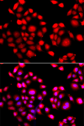
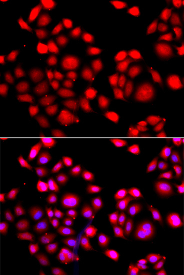
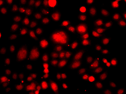
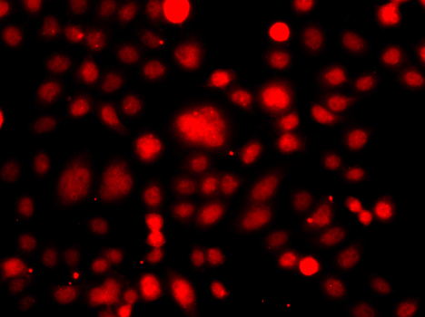
- Immunofluorescence analysis of A549 cell using PRDM14 antibody. Blue: DAPI for nuclear staining.
- Submitted by
- GeneTex (provider)
- Main image
- Experimental details
- Immunofluorescence analysis of A549 cell using PRDM14 antibody.
Supportive validation
- Submitted by
- GeneTex (provider)
- Main image
- Experimental details
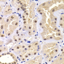
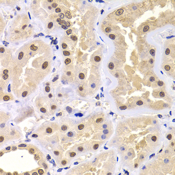
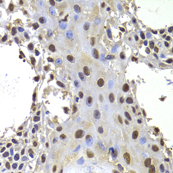
- Immunohistochemistry of paraffin-embedded human kidney using PRDM14 antibody at dilution of 1:100 (x400 lens).
- Submitted by
- GeneTex (provider)
- Main image

- Experimental details
- Immunohistochemistry of paraffin-embedded human well-differentiated squamous skin carcinoma using PRDM14 antibody at dilution of 1:100 (x400 lens).
Supportive validation
- Submitted by
- GeneTex (provider)
- Main image

- Experimental details
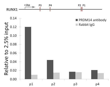
- ChIP analysis was performed with 293 lysates and either PRDM14 antibody or control RABBIT IgG. The precipitated DNA was detected by qPCR with primer sets targeting to P1, P2, P3 and P4 regions of RUNX1 gene as the schematic diagram illustrated. Histogram was constructed by the ratios of the immunoprecipitated DNA to the input.