Explore
 Validate
 Learn
Learn Western blot
Antibody data
- Antibody Data
- Antigen structure
- References [9]
- Comments [0]
- Validations
- Western blot [3]
- ELISA [1]
- Immunohistochemistry [1]
Submit
Validation data
Reference
Comment
Report error
- Product number
- AF2948 - Provider product page

- Provider
- R&D Systems
- Product name
- Mouse Serum Amyloid A1/A2 Antibody
- Antibody type
- Polyclonal
- Description
- Antigen Affinity-purified. Detects mouse Serum Amyloid A1/SAA1 and mouse Serum Amyloid A2/SAA2 in direct ELISAs and Western blots. In direct ELISAs, less than 3% cross-reactivity with recombinant human (rh) SAA1, rhSAA2, recombinant mouse (rm) SAA3, and rmSAA4 is observed.
- Reactivity
- Mouse
- Host
- Goat
- Conjugate
- Unconjugated
- Antigen sequence
P05366- Isotype
- IgG
- Vial size
- 100 ug
- Concentration
- LYOPH
- Storage
- Use a manual defrost freezer and avoid repeated freeze-thaw cycles. 12 months from date of receipt, -20 to -70 °C as supplied. 1 month, 2 to 8 °C under sterile conditions after reconstitution. 6 months, -20 to -70 °C under sterile conditions after reconstitution.
Submitted references Serum Amyloid A Protein as a Potential Biomarker for Severity and Acute Outcome in Traumatic Brain Injury.
Increased hypothalamic microglial activation after viral-induced pneumococcal lung infection is associated with excess serum amyloid A production.
Amyloid deposition in a mouse model humanized at the transthyretin and retinol-binding protein 4 loci.
Serum Amyloid A1 Is an Epithelial Prorestitutive Factor.
Scavenger Receptor A1 Prevents Metastasis of Non-Small Cell Lung Cancer via Suppression of Macrophage Serum Amyloid A1.
Zymosan-mediated inflammation impairs in vivo reverse cholesterol transport.
Transmission of circulating cell-free AA amyloid oligomers in exosomes vectors via a prion-like mechanism.
Role of APAF-1, E-cadherin and peritumoral lymphocytic infiltration in tumour budding in colorectal cancer.
Fish oil increases cholesterol storage in white adipose tissue with concomitant decreases in inflammation, hepatic steatosis, and atherosclerosis in mice.
Wicker E, Benton L, George K, Furlow W, Villapol S
BioMed research international 2019;2019:5967816
BioMed research international 2019;2019:5967816
Increased hypothalamic microglial activation after viral-induced pneumococcal lung infection is associated with excess serum amyloid A production.
Wang H, Blackall M, Sominsky L, Spencer SJ, Vlahos R, Churchill M, Bozinovski S
Journal of neuroinflammation 2018 Jul 6;15(1):200
Journal of neuroinflammation 2018 Jul 6;15(1):200
Amyloid deposition in a mouse model humanized at the transthyretin and retinol-binding protein 4 loci.
Li X, Lyu Y, Shen J, Mu Y, Qiang L, Liu L, Araki K, Imbimbo BP, Yamamura KI, Jin S, Li Z
Laboratory investigation; a journal of technical methods and pathology 2018 Apr;98(4):512-524
Laboratory investigation; a journal of technical methods and pathology 2018 Apr;98(4):512-524
Serum Amyloid A1 Is an Epithelial Prorestitutive Factor.
Hinrichs BH, Matthews JD, Siuda D, O'Leary MN, Wolfarth AA, Saeedi BJ, Nusrat A, Neish AS
The American journal of pathology 2018 Apr;188(4):937-949
The American journal of pathology 2018 Apr;188(4):937-949
Scavenger Receptor A1 Prevents Metastasis of Non-Small Cell Lung Cancer via Suppression of Macrophage Serum Amyloid A1.
Zhang Y, Wei Y, Jiang B, Chen L, Bai H, Zhu X, Li X, Zhang H, Yang Q, Ma J, Xu Y, Ben J, Christiani DC, Chen Q
Cancer research 2017 Apr 1;77(7):1586-1598
Cancer research 2017 Apr 1;77(7):1586-1598
Zymosan-mediated inflammation impairs in vivo reverse cholesterol transport.
Malik P, Berisha SZ, Santore J, Agatisa-Boyle C, Brubaker G, Smith JD
Journal of lipid research 2011 May;52(5):951-7
Journal of lipid research 2011 May;52(5):951-7
Transmission of circulating cell-free AA amyloid oligomers in exosomes vectors via a prion-like mechanism.
Tasaki M, Ueda M, Ochiai S, Tanabe Y, Murata S, Misumi Y, Su Y, Sun X, Shinriki S, Jono H, Shono M, Obayashi K, Ando Y
Biochemical and biophysical research communications 2010 Oct 1;400(4):559-62
Biochemical and biophysical research communications 2010 Oct 1;400(4):559-62
Role of APAF-1, E-cadherin and peritumoral lymphocytic infiltration in tumour budding in colorectal cancer.
Zlobec I, Lugli A, Baker K, Roth S, Minoo P, Hayashi S, Terracciano L, Jass JR
The Journal of pathology 2007 Jul;212(3):260-8
The Journal of pathology 2007 Jul;212(3):260-8
Fish oil increases cholesterol storage in white adipose tissue with concomitant decreases in inflammation, hepatic steatosis, and atherosclerosis in mice.
Saraswathi V, Gao L, Morrow JD, Chait A, Niswender KD, Hasty AH
The Journal of nutrition 2007 Jul;137(7):1776-82
The Journal of nutrition 2007 Jul;137(7):1776-82
No comments: Submit comment
Supportive validation
- Submitted by
- R&D Systems (provider)
- Main image
- Experimental details
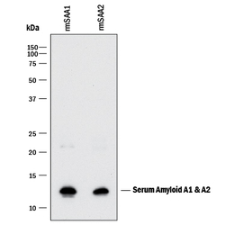
- Detection of Mouse Serum Amyloid A1/A2 by Western Blot. Western blot shows recombinant mouse Serum Amyloid A1 and recombinant mouse Serum Amyloid A2. PVDF membrane was probed with 1 µg/mL of Goat Anti-Mouse Serum Amyloid A1/A2 Antigen Affinity-purified Polyclonal Antibody (Catalog # AF2948) followed by HRP-conjugated Anti-Goat IgG Secondary Antibody (Catalog # HAF017). A specific band was detected for Serum Amyloid A1/A2 at approximately 12 kDa (as indicated). This experiment was conducted under reducing conditions and using Immunoblot Buffer Group 1.

- Submitted by
- R&D Systems (provider)
- Main image
- Experimental details
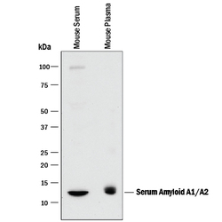
- Detection of Mouse Serum Amyloid A1/A2 by Western Blot. Western blot shows mouse serum and mouse plasma. PVDF membrane was probed with 1 µg/mL of Goat Anti-Mouse Serum Amyloid A1/A2 Antigen Affinity-purified Polyclonal Antibody (Catalog # AF2948) followed by HRP-conjugated Anti-Goat IgG Secondary Antibody (Catalog # HAF017). A specific band was detected for Serum Amyloid A1/A2 at approximately 12 kDa (as indicated). This experiment was conducted under reducing conditions and using Immunoblot Buffer Group 1.

- Submitted by
- R&D Systems (provider)
- Main image
- Experimental details
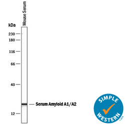
- Detection of Mouse Serum Amyloid A1/A2 by Simple WesternTM. Simple Western lane view shows mouse serum, loaded at a 1:100 dilution. A specific band was detected for Serum Amyloid A1/A2 at approximately 14 kDa (as indicated) using 50 µg/mL of (Catalog # AF2948) followed by 1:50 dilution of HRP-conjugated Anti-Goat IgG Secondary Antibody (Catalog # HAF109). This experiment was conducted under reducing conditions and using the 12-230 kDa separation system.

Supportive validation
- Submitted by
- R&D Systems (provider)
- Main image

- Experimental details
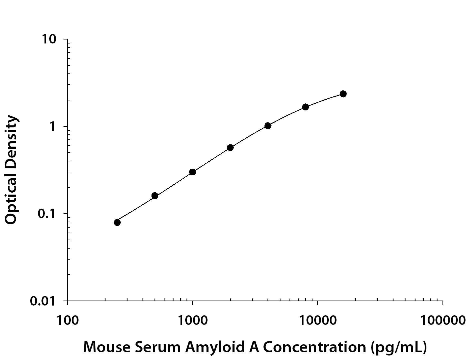
- Mouse Serum Amyloid A1/A2 ELISA Standard Curve. Recombinant Mouse Serum Amyloid A1/A2 protein was serially diluted 2-fold and captured by Rat Anti-Mouse Serum Amyloid A1/A2 Monoclonal Antibody (Catalog # MAB2948) coated on a Clear Polystyrene Microplate (Catalog # DY990). Goat Anti-Mouse Serum Amyloid A1/A2 Antigen Affinity-purified Polyclonal Antibody (Catalog # AF2948) was biotinylated and incubated with the protein captured on the plate. Detection of the standard curve was achieved by incubating Streptavidin-HRP (Catalog # DY998) followed by Substrate Solution (Catalog # DY999) and stopping the enzymatic reaction with Stop Solution (Catalog # DY994).
Supportive validation
- Submitted by
- R&D Systems (provider)
- Main image

- Experimental details
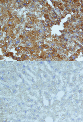
- Serum Amyloid A1/A2 in Mouse Liver. Serum Amyloid A1/A2 was detected in perfusion fixed frozen sections of mouse liver using Goat Anti-Mouse Serum Amyloid A1/A2 Antigen Affinity-purified Polyclonal Antibody (Catalog # AF2948) at 15 µg/mL overnight at 4 °C. Tissue was stained using the Anti-Goat HRP-DAB Cell & Tissue Staining Kit (brown; Catalog # CTS008) and counterstained with hematoxylin (blue). Lower panel shows a lack of labeling when primary antibodies are omitted and tissue is stained only with secondary antibody followed by incubation with detection reagents. Specific staining was localized to cytoplasm. View our protocol for Chromogenic IHC Staining of Frozen Tissue Sections.