 Explore
 Validate
 Learn
LearnSP5391
antibody from Acris Antibodies GmbH
Targeting: PDIA3
ERp57, ERp60, ERp61, GRP57, GRP58, HsT17083, P58, PI-PLC
 Western blot
 Immunoprecipitation
Antibody data
- Antibody Data
- Antigen structure
- References [1]
- Comments [0]
- Validations
- Western blot [2]
- Immunocytochemistry [3]
Submit
Validation data
Reference
Comment
Report error
- Product number
- SP5391 - Provider product page

- Provider
- Acris Antibodies GmbH
- Proper citation
- Acris Antibodies GmbH Cat#SP5391, RRID:AB_981020
- Product name
- anti PDIA3
- Antibody type
- Polyclonal
- Antigen
- Synthetic Peptide corresponding to residues 490-505 of Human ERp57
- Reactivity
- Human, Mouse, Rat, Bovine
- Host
- Rabbit
- Vial size
- 0.1 ml
Submitted references Tumor necrosis factor-α treatment of HepG2 cells mobilizes a cytoplasmic pool of ERp57/1,25D₃-MARRS to the nucleus.
Grindel BJ, Rohe B, Safford SE, Bennett JJ, Farach-Carson MC
Journal of cellular biochemistry 2011 Sep;112(9):2606-15
Journal of cellular biochemistry 2011 Sep;112(9):2606-15
No comments: Submit comment
Supportive validation
- Submitted by
- Acris Antibodies GmbH (provider)
- Main image

- Experimental details
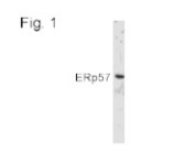
- Figure 1 shows a Western blot of ERp57 in HEK cell lysate using SP5391.
- Submitted by
- Acris Antibodies GmbH (provider)
- Main image
- Experimental details
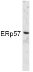
- Western blot of ERp29 in HEK cell lysate using PDIA3 Antibody Cat.-No SP5391

Supportive validation
- Submitted by
- Acris Antibodies GmbH (provider)
- Main image

- Experimental details
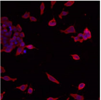
- Immunofluorescent analysis of ERp57 using polyclonal PDIA3 Antibody Cat.-No SP5391 shows staining in p19 Cells.
- Submitted by
- Acris Antibodies GmbH (provider)
- Main image
- Experimental details
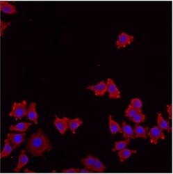
- Immunofluorescent analysis of ERp57 using polyclonal PDIA3 Antibody Cat.-No SP5391 shows staining in NS-1 Cells.

- Submitted by
- Acris Antibodies GmbH (provider)
- Main image
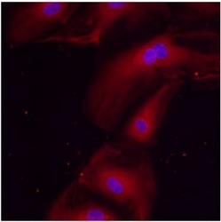
- Experimental details
- Immunofluorescent analysis of ERp57 using polyclonal PDIA3 Antibody Cat.-No SP5391 shows staining in HMVEC Cells.
