Explore
 Validate
 Learn
LearnHPA003230
antibody from Atlas Antibodies
Targeting: PDIA3
ERp57, ERp60, ERp61, GRP57, GRP58, HsT17083, P58, PI-PLC
 Western blot
Antibody data
- Antibody Data
- Antigen structure
- References [4]
- Comments [0]
- Validations
- Western blot [3]
- Immunocytochemistry [2]
- Immunohistochemistry [6]
Submit
Validation data
Reference
Comment
Report error
- Product number
- HPA003230 - Provider product page

- Provider
- Atlas Antibodies
- Proper citation
- Atlas Antibodies Cat#HPA003230, RRID:AB_1079030
- Product name
- Anti-PDIA3
- Antibody type
- Polyclonal
- Reactivity
- Human, Mouse, Rat
- Host
- Rabbit
- Conjugate
- Unconjugated
- Antigen sequence
PTLKIFRDGEEAGAYDGPRTADGIVSHLKKQAGPA
SVPLRTEEEFKKFISDKDASIVGFFDDSFSEAHSE
FLKAASNLRDNYRFAHTNVESLVNEYDDNGEGIIL
FRPSHLTNKFEDK- Isotype
- IgG
- Vial size
- 100 µl
- Storage
- Store at +4°C for short term storage. Long time storage is recommended at -20°C.
Submitted references The dehydrogenase region of the NADPH oxidase component Nox2 acts as a protein disulfide isomerase (PDI) resembling PDIA3 with a role in the binding of the activator protein p67 (phox.).
Proteomics based identification of cell migration related proteins in HBV expressing HepG2 cells.
Loss-of-function mutations in MICU1 cause a brain and muscle disorder linked to primary alterations in mitochondrial calcium signaling
The involvement of SMILE/TMTC3 in endoplasmic reticulum stress response.
Bechor E, Dahan I, Fradin T, Berdichevsky Y, Zahavi A, Federman Gross A, Rafalowski M, Pick E
Frontiers in chemistry 2015;3:3
Frontiers in chemistry 2015;3:3
Proteomics based identification of cell migration related proteins in HBV expressing HepG2 cells.
Feng H, Li X, Chan V, Chen WN
PloS one 2014;9(4):e95621
PloS one 2014;9(4):e95621
Loss-of-function mutations in MICU1 cause a brain and muscle disorder linked to primary alterations in mitochondrial calcium signaling
Logan C, Szabadkai G, Sharpe J, Parry D, Torelli S, Childs A, Kriek M, Phadke R, Johnson C, Roberts N, Bonthron D, Pysden K, Whyte T, Munteanu I, Foley A, Wheway G, Szymanska K, Natarajan S, Abdelhamed Z, Morgan J, Roper H, Santen G, Niks E, van der Pol W, Lindhout D, Raffaello A, De Stefani D, den Dunnen J, Sun Y, Ginjaar I, Sewry C, Hurles M, Rizzuto R, Duchen M, Muntoni F, Sheridan E
Nature Genetics 2013 December;46(2):188-193
Nature Genetics 2013 December;46(2):188-193
The involvement of SMILE/TMTC3 in endoplasmic reticulum stress response.
Racapé M, Duong Van Huyen JP, Danger R, Giral M, Bleicher F, Foucher Y, Pallier A, Pilet P, Tafelmeyer P, Ashton-Chess J, Dugast E, Pettré S, Charreau B, Soulillou JP, Brouard S
PloS one 2011;6(5):e19321
PloS one 2011;6(5):e19321
No comments: Submit comment
Enhanced validation
- Submitted by
- Atlas Antibodies (provider)
- Enhanced method
- Genetic validation
- Main image

- Experimental details
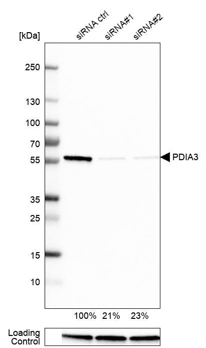
- Western blot analysis in U-251MG cells transfected with control siRNA, target specific siRNA probe #1 and #2, using Anti-PDIA3 antibody. Remaining relative intensity is presented. Loading control: Anti-GAPDH.
- Submitted by
- Atlas Antibodies (provider)
- Enhanced method
- Genetic validation
- Main image
- Experimental details
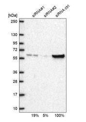
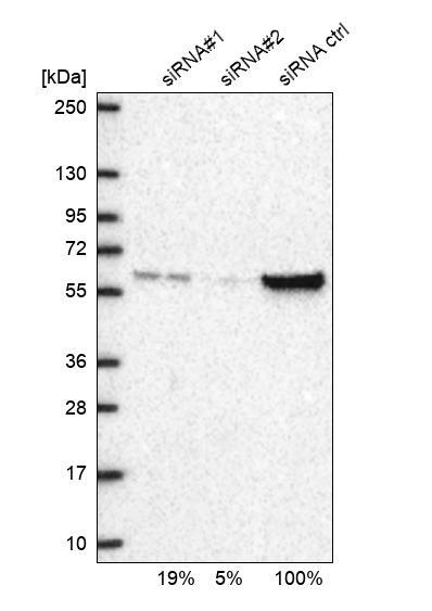
- Western blot analysis in U2OS cells transfected with control siRNA, target specific siRNA probe #1 and #2, using Anti-PDIA3 antibody. Remaining relative intensity is presented
- Submitted by
- Atlas Antibodies (provider)
- Enhanced method
- Independent antibody validation
- Main image
- Experimental details
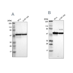
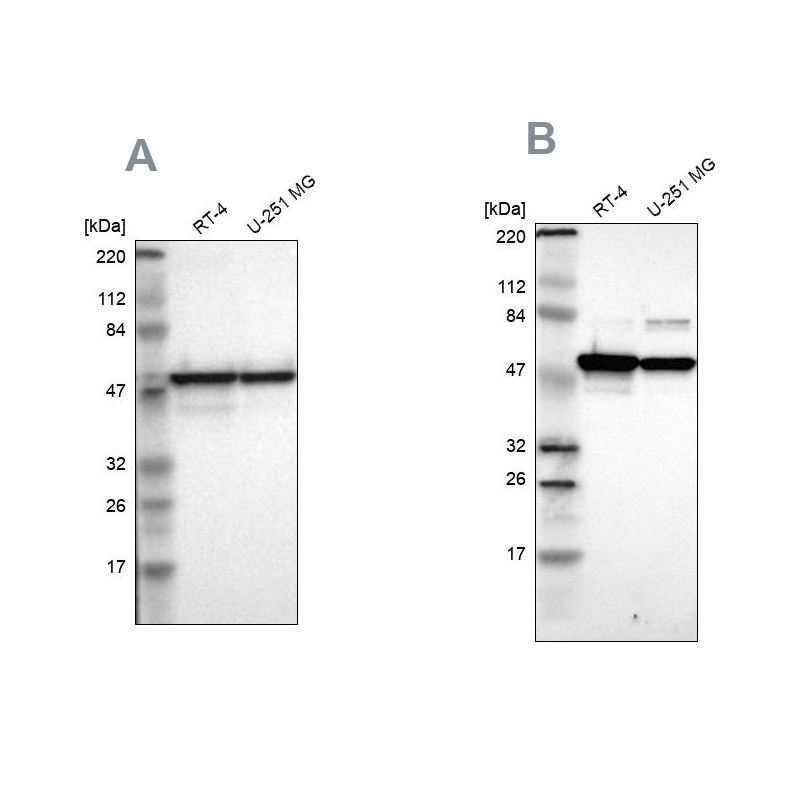
- Western blot analysis using Anti-PDIA3 antibody HPA003230 (A) shows similar pattern to independent antibody HPA002645 (B).
Enhanced validation
Supportive validation
- Submitted by
- 55af80e3e0991
- Enhanced method
- Genetic validation
- Main image
- Experimental details
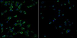
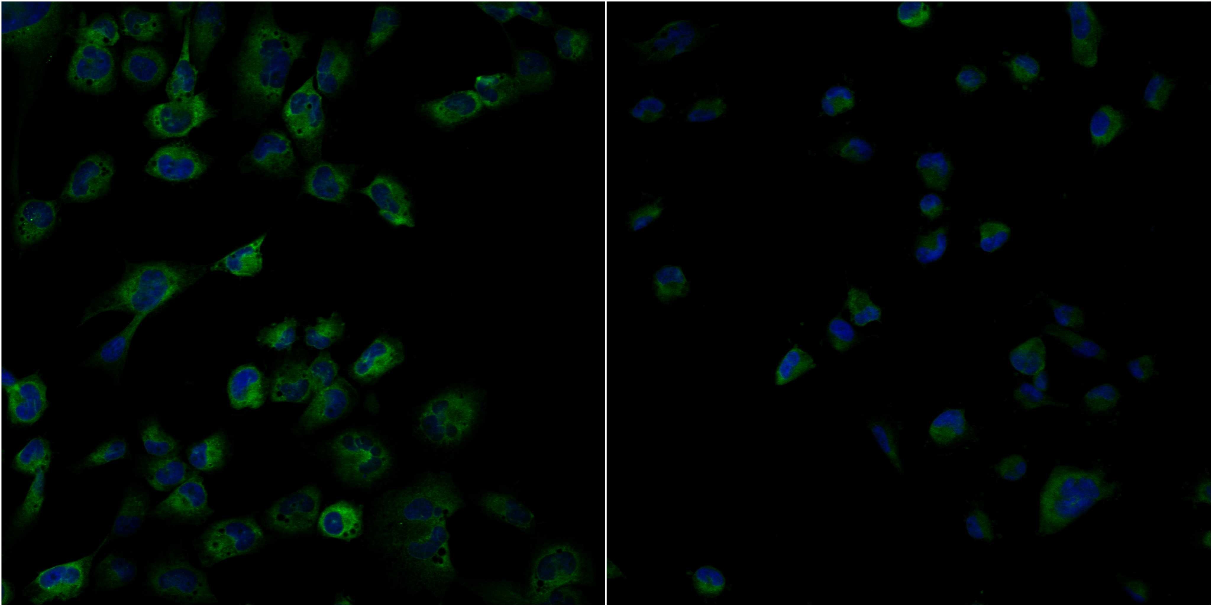
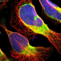
- Confocal images of immunofluorescently stained human U-2 OS cells.The protein PDIA3 is shown in green and the nucleus in blue. The image to the left show cells transfected with control siRNA and the image to the right show cells where PDIA3 has been downregulated with specific siRNA.
- Sample type
- U-2 OS cells
- Primary Ab dilution
- 1:127
- Secondary Ab
- Secondary Ab
- Secondary Ab dilution
- 1:800
- Knockdown/Genetic Approaches Application
- Immunocytochemistry
Supportive validation
- Submitted by
- Atlas Antibodies (provider)
- Main image
- Experimental details
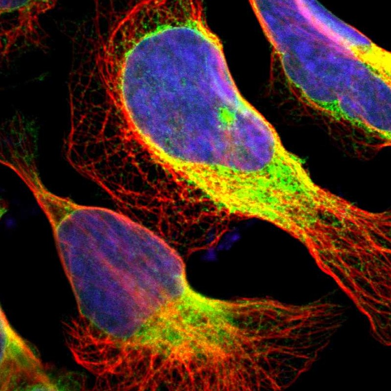
- Immunofluorescent staining of human cell line U-2 OS shows localization to endoplasmic reticulum.
- Sample type
- HUMAN
Enhanced validation
Enhanced validation
Supportive validation
- Submitted by
- Atlas Antibodies (provider)
- Enhanced method
- Orthogonal validation
- Main image
- Experimental details
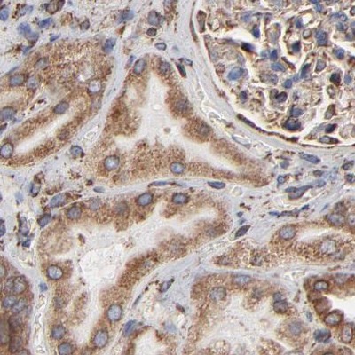
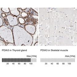
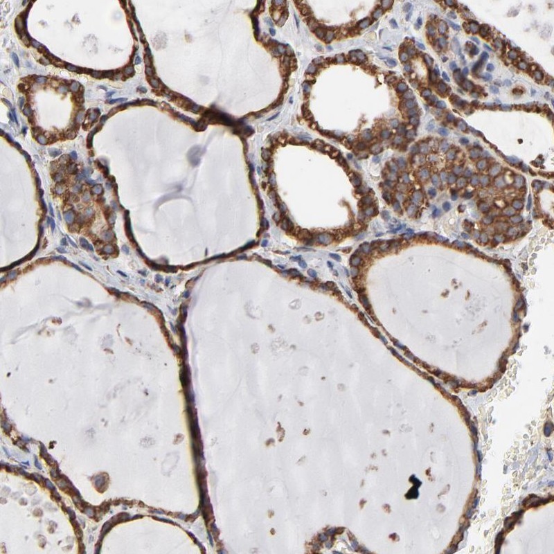
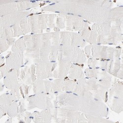
- Immunohistochemistry analysis in human thyroid gland and skeletal muscle tissues using Anti-PDIA3 antibody. Corresponding PDIA3 RNA-seq data are presented for the same tissues.
- Sample type
- HUMAN

Enhanced validation
- Submitted by
- Atlas Antibodies (provider)
- Enhanced method
- Independent antibody validation
- Main image

- Experimental details
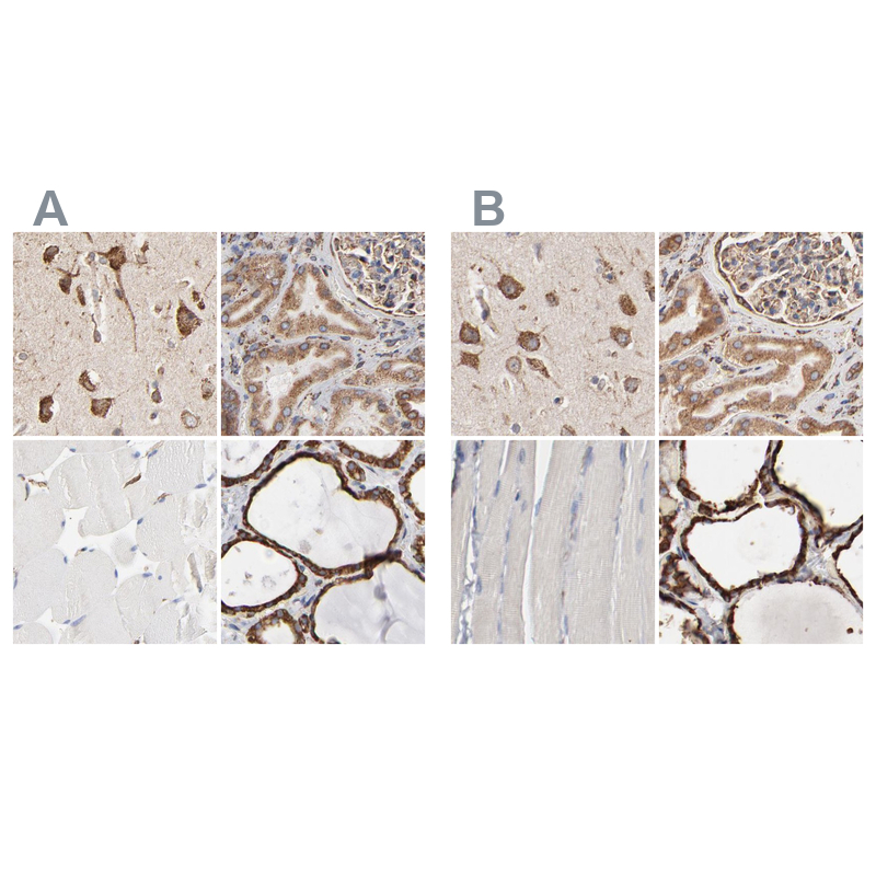
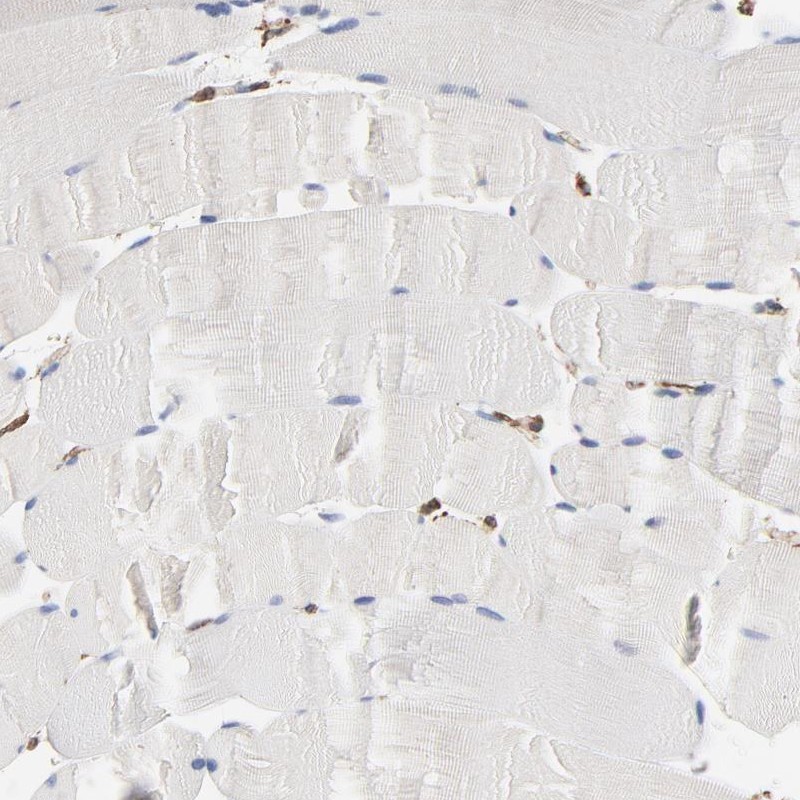
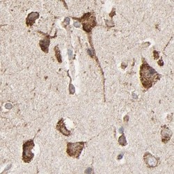
- Immunohistochemical staining of human cerebral cortex, kidney, skeletal muscle and thyroid gland using Anti-PDIA3 antibody HPA003230 (A) shows similar protein distribution across tissues to independent antibody HPA002645 (B).
Supportive validation
- Submitted by
- Atlas Antibodies (provider)
- Main image

- Experimental details
- Immunohistochemical staining of human thyroid gland shows high expression.
- Sample type
- HUMAN
- Submitted by
- Atlas Antibodies (provider)
- Main image
- Experimental details
- Immunohistochemical staining of human skeletal muscle shows low expression as expected.
- Sample type
- HUMAN
- Submitted by
- Atlas Antibodies (provider)
- Main image
- Experimental details
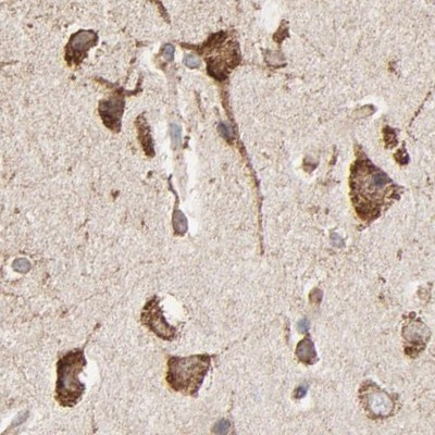
- Immunohistochemical staining of human cerebral cortex using Anti-PDIA3 antibody HPA003230.
- Sample type
- HUMAN
- Submitted by
- Atlas Antibodies (provider)
- Main image

- Experimental details
- Immunohistochemical staining of human kidney using Anti-PDIA3 antibody HPA003230.
- Sample type
- HUMAN