Explore
 Validate
 Learn
Learn Western blot
 ELISA
ELISAAntibody data
- Antibody Data
- Antigen structure
- References [2]
- Comments [0]
- Validations
- Western blot [3]
- Flow cytometry [2]
Submit
Validation data
Reference
Comment
Report error
- Product number
- NB110-68281 - Provider product page

- Provider
- Novus Biologicals
- Proper citation
- Novus Cat#NB110-68281, RRID:AB_1111093
- Product name
- Mouse Monoclonal TRF-1 Antibody
- Antibody type
- Monoclonal
- Description
- Protein G purified.
- Reactivity
- Human, Mouse, Rat, Simian
- Host
- Mouse
- Isotype
- IgG
- Vial size
- 0.1 ml
- Concentration
- 1.5 mg/ml
- Storage
- Store at 4C short term. Aliquot and store at -20C long term. Avoid freeze-thaw cycles.
Submitted references MicroRNA-214 modulates the senescence of vascular smooth muscle cells in carotid artery stenosis.
Eroded human telomeres are more prone to remain uncapped and to trigger a G2 checkpoint response.
Chen YL, Sheu JJ, Sun CK, Huang TH, Lin YP, Yip HK
Molecular medicine (Cambridge, Mass.) 2020 May 14;26(1):46
Molecular medicine (Cambridge, Mass.) 2020 May 14;26(1):46
Eroded human telomeres are more prone to remain uncapped and to trigger a G2 checkpoint response.
Jullien L, Mestre M, Roux P, Gire V
Nucleic acids research 2013 Jan;41(2):900-11
Nucleic acids research 2013 Jan;41(2):900-11
No comments: Submit comment
Supportive validation
- Submitted by
- Novus Biologicals (provider)
- Main image
- Experimental details
- Simple Western: TRF-1 Antibody (57-6) [NB110-68281] - Image shows a specific band for TRF1 in 0.5 mg/mL of HeLa lysate. This experiment was performed under reducing conditions using the 12-230 kDa separation system.
- Submitted by
- Novus Biologicals (provider)
- Main image
- Experimental details
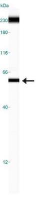
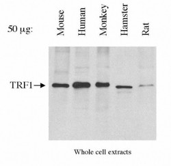
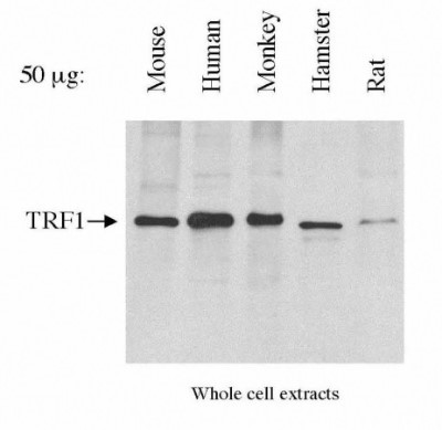
- Western Blot: TRF-1 Antibody (57-6) [NB110-68281] - Detection of TRF1. 50 ug of total lysate each lane.
- Submitted by
- Novus Biologicals (provider)
- Main image
- Experimental details
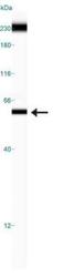
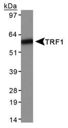
- Western Blot: TRF-1 Antibody (57-6) [NB110-68281] - Detection of TRF1 in HeLa whole cell extracts.

Supportive validation
- Submitted by
- Novus Biologicals (provider)
- Main image
- Experimental details
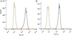
- Flow Cytometry: TRF-1 Antibody (57-6) [NB110-68281] - Intracellular flow cytometric staining of 1 x 10^6 CHO (A) and HEK-293 (B) cells using TRF1 antibody (dark blue). Isotype control shown in orange. An antibody concentration of 1 ug/1x10^6 cells was used.
- Submitted by
- Novus Biologicals (provider)
- Main image
- Experimental details
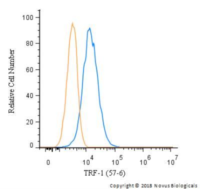
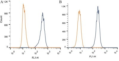
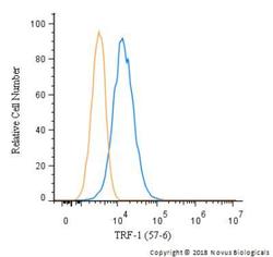
- Flow Cytometry: TRF-1 Antibody (57-6) [NB110-68281] - An intracellular stain was performed on HeLa with NB110-68281 (blue) and a matched isotype control (orange). Cells were fixed with 4% PFA and then permeablized with 0.1% saponin. Cells were incubated in an antibody dilution of 2.5 ug/mL for 30 minutes at room temperature, followed by mouse F(ab)2 IgG (H+L) APC-conjugated secondary antibody (F0101B, R&D Systems).