Explore
 Validate
 Learn
Learn Western blot
Antibody data
- Antibody Data
- Antigen structure
- References [0]
- Comments [0]
- Validations
- Western blot [5]
- Immunocytochemistry [1]
Submit
Validation data
Reference
Comment
Report error
- Product number
- PA5-47552 - Provider product page

- Provider
- Invitrogen Antibodies
- Product name
- MAFF Polyclonal Antibody
- Antibody type
- Polyclonal
- Antigen
- Recombinant full-length protein
- Description
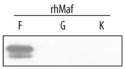
- In Western blots, less than 1% cross-reactivity with recombinant human (rh) MafG and rhMafK is observed.
- Concentration
- 0.2 mg/mL
No comments: Submit comment
Supportive validation
- Submitted by
- Invitrogen Antibodies (provider)
- Main image
- Experimental details
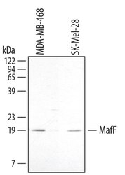
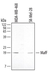
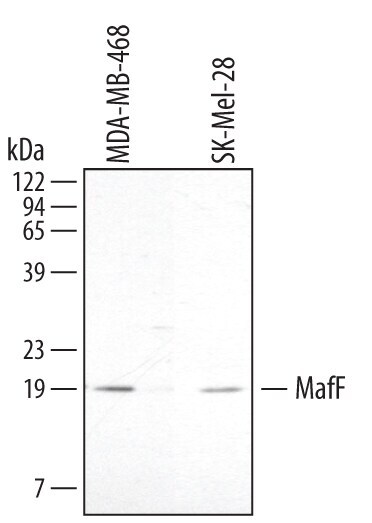
- Western blot analysis from lysates of MDA-MB-468 human breast cancer cell line and SK-Mel-28 human malignant melanoma cell line. PVDF membrane was probed with 2 µg/mL Goat Anti-human MafF Antigen Affinity-purified Polyclonal Antibody (Product # PA5-47552) followed by HRP-conjugated Anti-Goat IgG Secondary Antibody. A specific band for MafF was detected at approximately 19 kDa (as indicated). This experiment was conducted under reducing conditions.
- Submitted by
- Invitrogen Antibodies (provider)
- Main image
- Experimental details
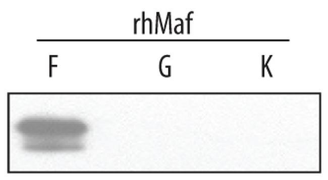
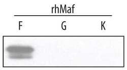
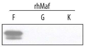
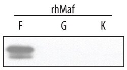
- Western blot analysis of MAFF in 2 ng/lane recombinant human (rh) MafF, MafG, and MafK. Samples were incubated in MAFF polyclonal antibody (Product # PA5-47552) using a dilution of 2 µg/mL followed by a HRP-conjugated Anti-Goat IgG secondary antibody. A specific band for MafF was detected at approximately 19 kDa (as indicated). This experiment was conducted under reducing conditions.
- Submitted by
- Invitrogen Antibodies (provider)
- Main image
- Experimental details
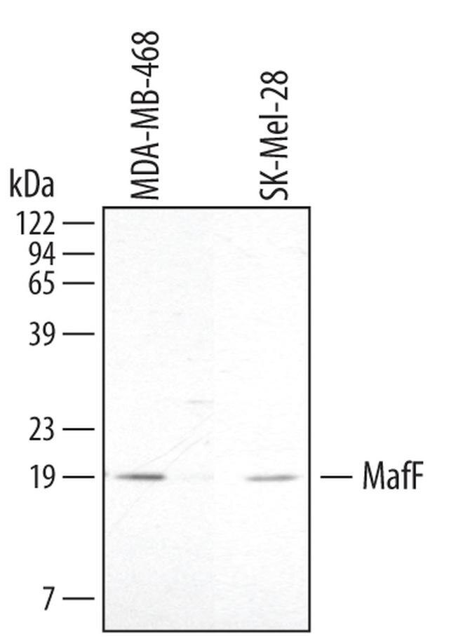
- Western blot analysis of MAFF in MDA-MB-468 human breast cancer cell line and SK-Mel-28 human malignant melanoma cell line. Samples were incubated in MAFF polyclonal antibody (Product # PA5-47552) using a dilution of 2 µg/mL followed by a HRP-conjugated Anti-Goat IgG secondary antibody. A specific band for MafF was detected at approximately 19 kDa (as indicated). This experiment was conducted under reducing conditions.
- Submitted by
- Invitrogen Antibodies (provider)
- Main image
- Experimental details
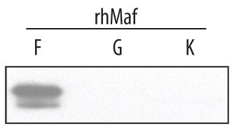
- Western blot analysis of MAFF in 2 ng/lane recombinant human (rh) MafF, MafG, and MafK. Samples were incubated in MAFF polyclonal antibody (Product # PA5-47552) using a dilution of 2 µg/mL followed by a HRP-conjugated Anti-Goat IgG secondary antibody. A specific band for MafF was detected at approximately 19 kDa (as indicated). This experiment was conducted under reducing conditions.
Supportive validation
- Submitted by
- Invitrogen Antibodies (provider)
- Main image
- Experimental details
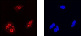
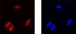
- Immunocytochemistry analysis of MAFF in immersion fixed HepG2 human hepatocellular carcinoma cell line. Samples were incubated in MAFF polyclonal antibody (Product # PA5-47552) using a dilution of 15 µg/mL for 3 hours at room temperature followed by NorthernLights™ 557-conjugated Anti-Goat IgG Secondary Antibody (left panel, red) and counterstained with DAPI (right panel, blue). Specific staining was localized to nuclei.