Explore
 Validate
 Learn
Learn Western blot
Antibody data
- Antibody Data
- Antigen structure
- References [2]
- Comments [0]
- Validations
- Western blot [4]
- Immunocytochemistry [1]
- Immunohistochemistry [2]
Submit
Validation data
Reference
Comment
Report error
- Product number
- GTX105124 - Provider product page

- Provider
- GeneTex
- Proper citation
- GeneTex Cat#GTX105124, RRID:AB_1951054
- Product name
- OGDH antibody [C2C3], C-term
- Antibody type
- Polyclonal
- Reactivity
- Human, Mouse
- Host
- Rabbit
Submitted references Transketolase Regulates the Metabolic Switch to Control Breast Cancer Cell Metastasis via the α-Ketoglutarate Signaling Pathway.
α-ketoglutarate dehydrogenase inhibition counteracts breast cancer-associated lung metastasis.
Tseng CW, Kuo WH, Chan SH, Chan HL, Chang KJ, Wang LH
Cancer research 2018 Jun 1;78(11):2799-2812
Cancer research 2018 Jun 1;78(11):2799-2812
α-ketoglutarate dehydrogenase inhibition counteracts breast cancer-associated lung metastasis.
Atlante S, Visintin A, Marini E, Savoia M, Dianzani C, Giorgis M, Sürün D, Maione F, Schnütgen F, Farsetti A, Zeiher AM, Bertinaria M, Giraudo E, Spallotta F, Cencioni C, Gaetano C
Cell death & disease 2018 Jul 9;9(7):756
Cell death & disease 2018 Jul 9;9(7):756
No comments: Submit comment
Supportive validation
- Submitted by
- GeneTex (provider)
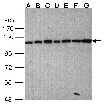
- Main image

- Experimental details
- Sample(30?g whole cell lysate)A: 293TB: A431 (GTX27909)C: H1299D: HeLa S3 (GTX14654)E: Hep G2 (GTX27900)F: MOLT4 (GTX27912)G: Raji (GTX27908)7.5% SDS PAGEGTX105124 diluted at 1:1000
- Validation comment
- WB
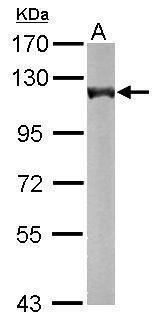
- Submitted by
- GeneTex (provider)
- Main image

- Experimental details
- Sample (50 ?g of whole cell lysate) A: Mouse brain 7.5% SDS PAGE GTX105124 diluted at 1:5000 The HRP-conjugated anti-rabbit IgG antibody (GTX213110-01) was used to detect the primary antibody.
- Submitted by
- GeneTex (provider)
- Main image
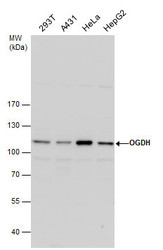
- Experimental details
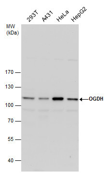
- OGDH antibody detects OGDH protein by western blot analysis. Various whole cell extracts (30 ?g) were separated by 7.5% SDS-PAGE, and blotted with OGDH antibody (GTX105124) diluted by 1:1000. The HRP-conjugated anti-rabbit IgG antibody (GTX213110-01) was used to detect the primary antibody.
- Submitted by
- GeneTex (provider)
- Main image
- Experimental details
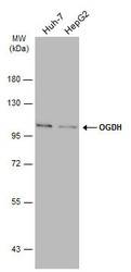
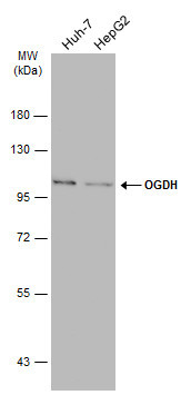
- Various whole cell extracts (30 ?g) were separated by 7.5% SDS-PAGE, and the membrane was blotted with OGDH antibody [C2C3], C-term (GTX105124) diluted at 1:1000. The HRP-conjugated anti-rabbit IgG antibody (GTX213110-01) was used to detect the primary antibody.
Supportive validation
- Submitted by
- GeneTex (provider)
- Main image
- Experimental details
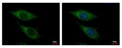
- OGDH antibody [C2C3], C-term detects OGDH protein at Mitochondria by immunofluorescent analysis. Sample: HeLa cells were fixed in -20¢J 100% MeOH for 5 min.Green: OGDH protein stained by OGDH antibody [C2C3], C-term (GTX105124) diluted at 1:500.Blue: Hoechst 33343 staining.

Supportive validation
- Submitted by
- GeneTex (provider)
- Main image
- Experimental details
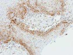
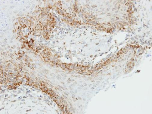
- Immunohistochemical analysis of paraffin-embedded Cal27 xenograft, using OGDH(GTX105124) antibody at 1:500 dilution.
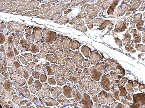
- Submitted by
- GeneTex (provider)
- Main image
- Experimental details
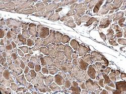
- OGDH antibody [C2C3], C-term detects OGDH protein at mitochondria on mouse muscle by immunohistochemical analysis. Sample: Paraffin-embedded mouse muscle. OGDH antibody [C2C3], C-term (GTX105124) dilution: 1:500.