Explore
 Validate
 Learn
Learn Western blot
Antibody data
- Antibody Data
- Antigen structure
- References [0]
- Comments [0]
- Validations
- Western blot [6]
- Immunocytochemistry [2]
- Immunohistochemistry [1]
Submit
Validation data
Reference
Comment
Report error
- Product number
- PA5-28089 - Provider product page

- Provider
- Invitrogen Antibodies
- Product name
- Cyclophilin A Polyclonal Antibody
- Antibody type
- Polyclonal
- Antigen
- Synthetic peptide
- Description
- Recommended positive controls: HepG2, Raji WCE, Raji C+M, Raji NE, mouse brain.
- Concentration
- 1 mg/mL
No comments: Submit comment
Supportive validation
- Submitted by
- Invitrogen Antibodies (provider)
- Main image
- Experimental details
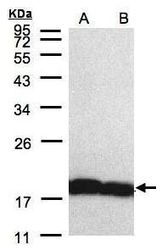
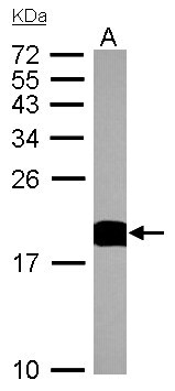
- Western blot analysis of Cyclophilin A using 30 µg of A) H1299 and B) HeLa S3 lysate. Samples were loaded onto a 12% SDS-PAGE gel and probed with a Cyclophilin A polyclonal antibody (Product # PA5-28089) at a dilution of 1:5000.
- Submitted by
- Invitrogen Antibodies (provider)
- Main image
- Experimental details
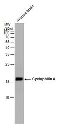
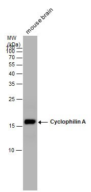
- Western Blot analysis of Cyclophilin A was performed by separating 50 µg of mouse tissue extracts by 15% SDS-PAGE. Proteins were transferred to a membrane and probed with a Cyclophilin A Polyclonal Antibody (Product # PA5-28089) at a dilution of 1:20000.
- Submitted by
- Invitrogen Antibodies (provider)
- Main image
- Experimental details
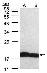
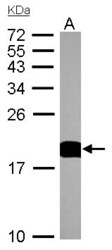
- Western Blot analysis of Cyclophilin A was performed by separating 30 µg of Hep G2 lysates by 12% SDS PAGE. Proteins were transferred to a membrane and probed with a Cyclophilin A Polyclonal Antibody (Product # PA5-28089) at a dilution of 1:3000.
- Submitted by
- Invitrogen Antibodies (provider)
- Main image
- Experimental details
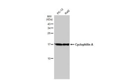
- Western Blot analysis of Cyclophilin A was performed by separating 30 µg of various whole cell extracts by 15% SDS-PAGE. Proteins were transferred to a membrane and probed with a Cyclophilin A Polyclonal Antibody (Product # PA5-28089) at a dilution of 1:10000 and a HRP-conjugated anti-rabbit IgG secondary antibody.

- Submitted by
- Invitrogen Antibodies (provider)
- Main image
- Experimental details
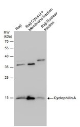
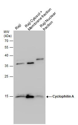
- Cyclophilin A antibody detects Cyclophilin A protein by western blot analysis. Raji whole cell extracts and cytoplasma+membrane and nuclear extracts (30 µg) were separated by 12% SDS-PAGE, and the membrane was blotted with Cyclophilin A antibody Cyclophilin A Polyclonal Antibody (Product # PA5-28089) at a dilution of 1:5,000. The HRP-conjugated anti-rabbit IgG antibody was used to detect the primary antibody.
- Submitted by
- Invitrogen Antibodies (provider)
- Main image
- Experimental details
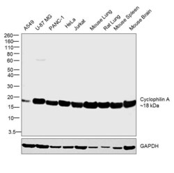
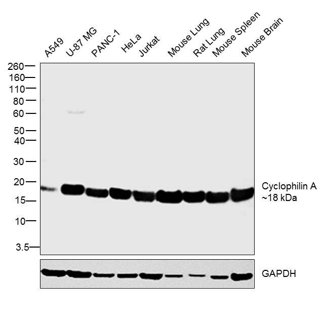
- Western blot was performed using Anti-Cyclophilin A Polyclonal Antibody (Product # PA5-28089) and a 18 kDa band corresponding to Cyclophilin A was observed across the cell lines and tissues tested. Whole cell extracts (30ug lysate) of A549 (Lane 1), U-87 MG (Lane 2), PANC-1 (Lane 3) , HeLa (Lane 4) , Jurkat (Lane 5), Mouse Lung (Lane 6), Rat Lung (Lane 7), Mouse Spleen (Lane 8) and Mouse Brain (Lane 9) were electrophoresed using Novex® NuPAGE® 4-12 % Bis-Tris gel (Product # NP0322BOX). Resolved proteins were then transferred onto a nitrocellulose membrane (Product # IB23001) by iBlot® 2 Dry Blotting System (Product # IB21001). The blot was probed with the primary antibody (1:5000 dilution) and detected by chemiluminescence with Goat anti-Rabbit IgG (H+L) Superclonal™ Recombinant Secondary Antibody, HRP (Product # A27036, 1:4000 dilution) using the iBright FL 1000 (Product # A32752). Chemiluminescent detection was performed using Novex® ECL Chemiluminescent Substrate Reagent Kit (Product # WP20005).
Supportive validation
- Submitted by
- Invitrogen Antibodies (provider)
- Main image

- Experimental details
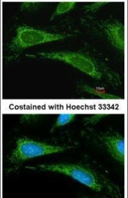
- Immunofluorescent analysis of Cyclophilin an In methanol-fixed HeLa cells using a Cyclophilin A polyclonal antibody (Product # PA5-28089) at a 1:200 dilution.
- Submitted by
- Invitrogen Antibodies (provider)
- Main image
- Experimental details
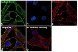
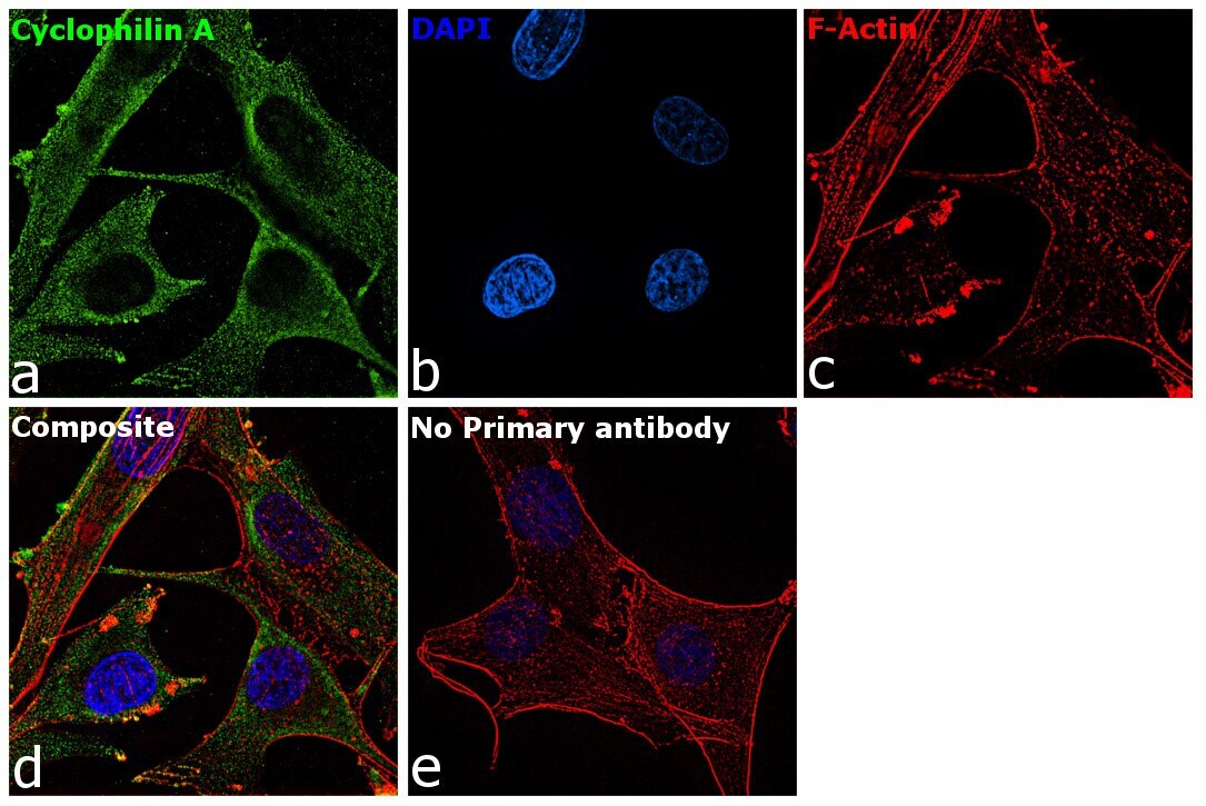
- Immunofluorescence analysis of Cyclophilin A was performed using 70% confluent log phase U-87 MG cells. The cells were fixed with 4% paraformaldehyde for 10 minutes, permeabilized with 0.1% Triton™ X-100 for 15 minutes, and blocked with 1% BSA for 1 hour at room temperature. The cells were labeled with Cyclophilin A Rabbit Polyclonal Antibody (Product # PA5-28089) at 1:200 dilution in 0.1% BSA and incubated overnight at 4 degree and then labeled with Goat anti-Rabbit IgG (H+L) Superclonal™ Recombinant Secondary Antibody, Alexa Fluor® 488 conjugate (Product # A27034) at a dilution of 1:2000 for 45 minutes at room temperature (Panel a: green). Nuclei (Panel b: blue) were stained with ProLong™ Diamond Antifade Mountant with DAPI (Product # P36962). F-actin (Panel c: red) was stained with Rhodamine Phalloidin (Product # R415, 1:300). Panel d represents the composite image showing cytoplasmic localization of Cyclophilin A. Panel e represents control cells with no primary antibody to assess background. The images were captured at 60X magnification.
Supportive validation
- Submitted by
- Invitrogen Antibodies (provider)
- Main image
- Experimental details
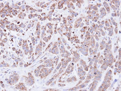
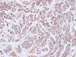
- Immunohistochemical analysis of paraffin-embedded MDA-MB-468 xenograft, using Cyclophilin A (Product # PA5-28089) antibody at 1:500 dilution. Antigen Retrieval: EDTA based buffer, pH 8.0, 15 min.