 Explore
 Validate
 Learn
Learn Immunocytochemistry
Antibody data
- Antibody Data
- Antigen structure
- References [0]
- Comments [0]
- Validations
- Immunocytochemistry [5]
- Immunohistochemistry [4]
Submit
Validation data
Reference
Comment
Report error
- Product number
- AMAb90992 - Provider product page

- Provider
- Atlas Antibodies
- Proper citation
- Atlas Antibodies Cat#AMAb90992, RRID:AB_2665753
- Product name
- Anti-ZYX
- Antibody type
- Monoclonal
- Reactivity
- Human
- Host
- Mouse
- Conjugate
- Unconjugated
- Antigen sequence
PAPKFSPVTPKFTPVASKFSPGAPGGSGSQPNQKL
GHPEALSAGTGSPQPPSFTYAQQREKPRVQEKQHP
VPPPAQNQNQVRSPGAPGPLTLKEVEELEQLTQQL
MQDMEHPQRQNVAVNE- Epitope
- Binds to an epitope located within the peptide sequence APGPLTLKEVEELEQ as determined by overlapping synthetic peptides.
- Isotype
- IgG
- Antibody clone number
- CL2502
- Vial size
- 100 µl
- Storage
- Store at +4°C for short term storage. Long time storage is recommended at -20°C.
No comments: Submit comment
Supportive validation
- Submitted by
- Atlas Antibodies (provider)
- Main image

- Experimental details
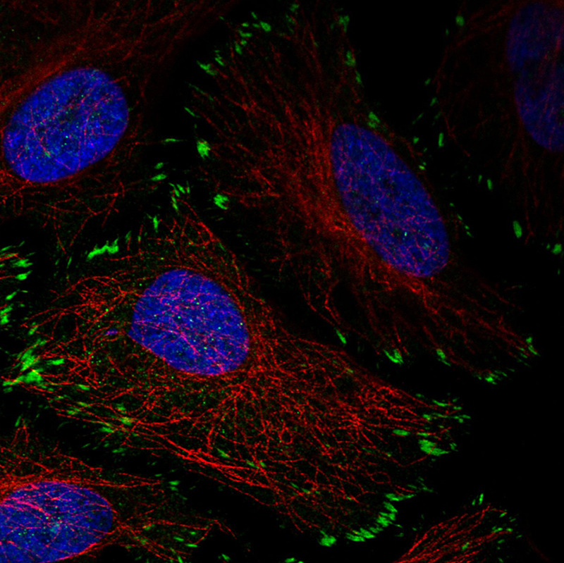
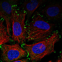
- Immunofluorescence staining in HeLa cell line with Anti-ZYX monoclonal antibody, showing distinct focal adhesion staining in green. Microtubule- and nuclear probes are visualized in red and blue respectively (where available).
- Sample type
- HUMAN
- Submitted by
- Atlas Antibodies (provider)
- Main image
- Experimental details
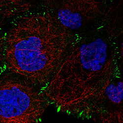
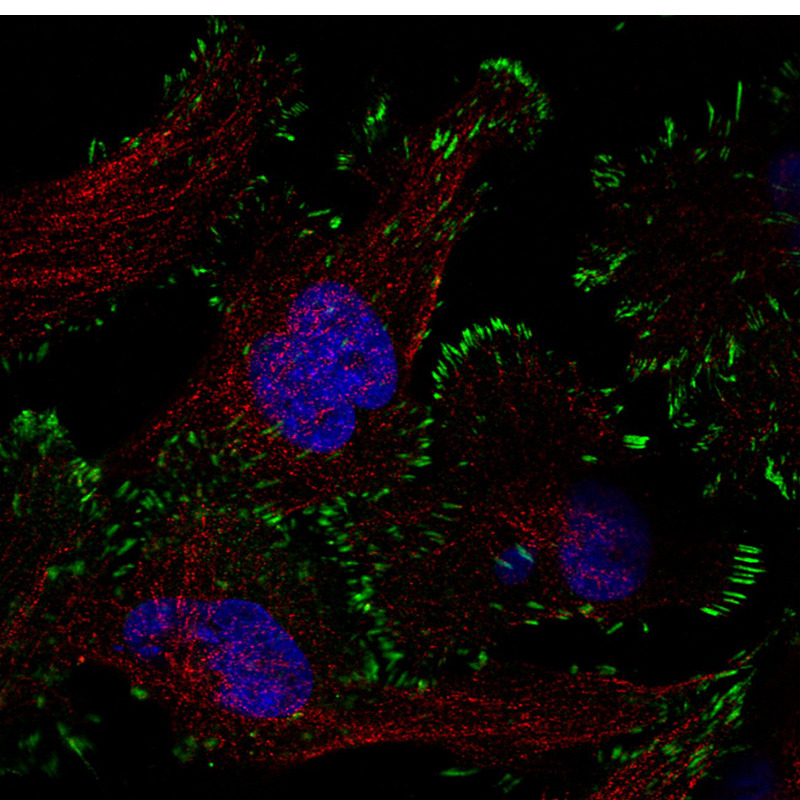
- Immunofluorescence staining in A431 cell line with Anti-ZYX monoclonal antibody, showing distinct focal adhesion staining in green. Microtubule- and nuclear probes are visualized in red and blue respectively (where available).
- Sample type
- HUMAN
- Submitted by
- Atlas Antibodies (provider)
- Main image
- Experimental details
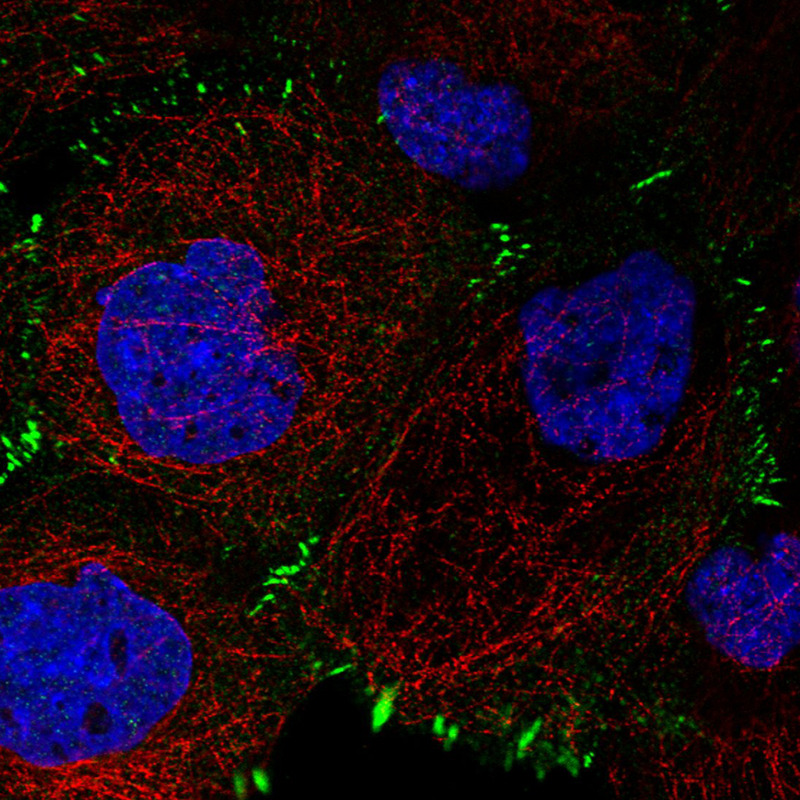
- Immunofluorescence staining in MCF7 cell line with Anti-ZYX monoclonal antibody, showing distinct focal adhesion staining in green. Microtubule- and nuclear probes are visualized in red and blue respectively (where available).
- Sample type
- HUMAN

- Submitted by
- Atlas Antibodies (provider)
- Main image
- Experimental details
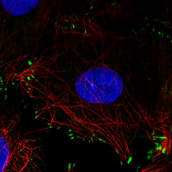
- Immunofluorescence staining in U2OS cell line with Anti-ZYX monoclonal antibody, showing distinct focal adhesion staining in green. Microtubule- and nuclear probes are visualized in red and blue respectively (where available).
- Sample type
- HUMAN

- Submitted by
- Atlas Antibodies (provider)
- Main image

- Experimental details
- Immunofluorescence staining in U251 cell line with Anti-ZYX monoclonal antibody, showing distinct focal adhesion staining in green. Microtubule- and nuclear probes are visualized in red and blue respectively (where available).
- Sample type
- HUMAN
Supportive validation
- Submitted by
- Atlas Antibodies (provider)
- Main image
- Experimental details
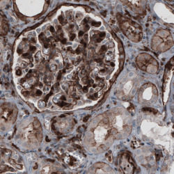
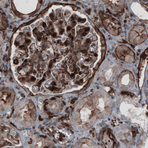
- Immunohistochemical staining of human kidney shows strong membranous and moderate cytoplasmic immunoreactivity in renal glomeruli and tubuli.
- Submitted by
- Atlas Antibodies (provider)
- Main image
- Experimental details
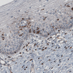
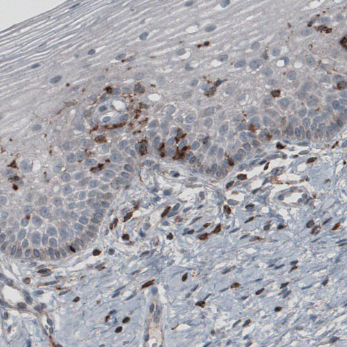
- Immunohistochemical staining of human skin shows strong cytoplasmic and membranous positivity in fibroblasts.
- Submitted by
- Atlas Antibodies (provider)
- Main image
- Experimental details
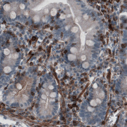
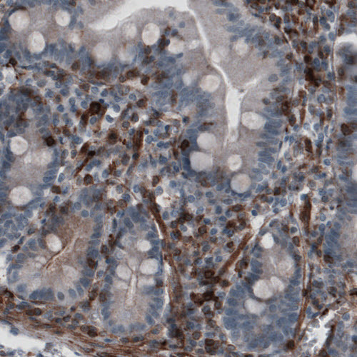
- Immunohistochemical staining of human small intestine shows cytoplasmic and membranous immunoreactivity in lymphoid and smooth muscle cells.
- Submitted by
- Atlas Antibodies (provider)
- Main image
- Experimental details
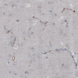
- Immunohistochemical staining of human cerebral cortex shows absence of immunoreactivity in neuronal cells (negative control).
