Explore
 Validate
 Learn
Learn Western blot
 Immunoprecipitation
Antibody data
- Antibody Data
- Antigen structure
- References [2]
- Comments [0]
- Validations
- Western blot [1]
- Immunocytochemistry [1]
Submit
Validation data
Reference
Comment
Report error
- Product number
- MA1-12300 - Provider product page

- Provider
- Invitrogen Antibodies
- Product name
- CDK7 Monoclonal Antibody (MO-1)
- Antibody type
- Monoclonal
- Antigen
- Recombinant protein fragment
- Reactivity
- Human
- Host
- Mouse
- Isotype
- IgG
- Antibody clone number
- MO-1
- Vial size
- 100 µg
- Concentration
- 1 mg/mL
- Storage
- -20° C, Avoid Freeze/Thaw Cycles
Submitted references The role of XPD in cell apoptosis and viability and its relationship with p53 and cdk2 in hepatoma cells.
Application of laser capture microdissection and differential display technique for screening of pathogenic genes involved in endometrial carcinoma.
Wang HY, Xiong GF, Zhang JX, Xu H, Guo WH, Xu JJ, Xiong XY
Medical oncology (Northwood, London, England) 2012 Mar;29(1):161-7
Medical oncology (Northwood, London, England) 2012 Mar;29(1):161-7
Application of laser capture microdissection and differential display technique for screening of pathogenic genes involved in endometrial carcinoma.
Wen-Xin L, Xi-Shan H
International journal of gynecological cancer : official journal of the International Gynecological Cancer Society 2007 Nov-Dec;17(6):1224-30
International journal of gynecological cancer : official journal of the International Gynecological Cancer Society 2007 Nov-Dec;17(6):1224-30
No comments: Submit comment
Supportive validation
- Submitted by
- Invitrogen Antibodies (provider)
- Main image
- Experimental details
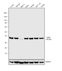
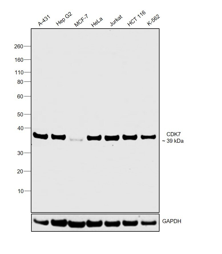
- Western blot was performed using Anti-CDK7 Monoclonal Antibody (MO-1) (Product # MA1-12300) and a 39 kDa band corresponding to CDK7 was observed across cell lines tested. Whole cell extracts (30 ug lysate) of A-431 (Lane 1), Hep G2 (Lane 2), MCF-7 (Lane 3), HeLa (Lane 4), Jurkat (Lane 5), HCT116 (Lane 6), K-562 (Lane 7) were electrophoresed using NuPAGE™ 4-12% Bis-Tris Protein Gel (Product # NP0322BOX). Resolved proteins were then transferred onto a nitrocellulose membrane (Product # IB23001) by iBlot® 2 Dry Blotting System (Product # IB21001). The blot was probed with the primary antibody (1:1000 dilution) and detected by chemiluminescence with Goat anti-Mouse IgG (H+L) Superclonal™ Recombinant Secondary Antibody, HRP (Product # A28177, 1:4000 dilution) using the iBright FL 1000 (Product # A32752). Chemiluminescent detection was performed using Novex® ECL Chemiluminescent Substrate Reagent Kit (Product # WP20005).
Supportive validation
- Submitted by
- Invitrogen Antibodies (provider)
- Main image
- Experimental details
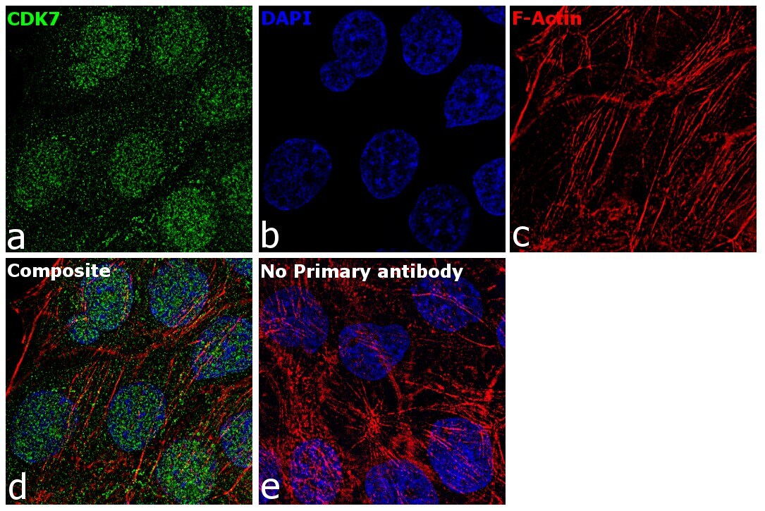
- Immunofluorescence analysis of CDK7 was performed using 70% confluent log phase A-431 cells. The cells were fixed with 4% paraformaldehyde for 10 minutes, permeabilized with 0.1% Triton™ X-100 for 15 minutes, and blocked with 2% BSA for 1 hour at room temperature. The cells were labeled with CDK7 Mouse Monoclonal Antibody (MO-1) (Product # MA1-12300) at 1:100 dilution in 0.1% BSA, incubated at 4 degree celsius overnight and then with Goat anti-Mouse IgG (H+L) Superclonal™ Recombinant Secondary Antibody, Alexa Fluor® 488 conjugate (Product # A28175) at a dilution of 1:2000 for 45 minutes at room temperature (Panel a: green). Nuclei (Panel b: blue) were stained with ProLong™ Diamond Antifade Mountant with DAPI (Product # P36962). F-actin (Panel c: red) was stained with Rhodamine Phalloidin (Product # R415, 1:300). Panel d represents the merged image showing nucleus and cytoplasmic localization. Panel e represents control cells with no primary antibody to assess background. The images were captured at 60X magnification.