Explore
 Validate
 Learn
Learn Western blot
 ELISA
ELISAAntibody data
- Antibody Data
- Antigen structure
- References [4]
- Comments [0]
- Validations
- Western blot [1]
- Immunocytochemistry [1]
- Immunohistochemistry [2]
Submit
Validation data
Reference
Comment
Report error
- Product number
- 14306-1-AP - Provider product page

- Provider
- Proteintech Group
- Proper citation
- Proteintech Cat#14306-1-AP, RRID:AB_10638478
- Product name
- WASL antibody
- Antibody type
- Polyclonal
- Description
- WASL antibody (Cat. #14306-1-AP) is a rabbit polyclonal antibody that shows reactivity with human, mouse, rat and has been validated for the following applications: IF, IHC, WB,ELISA.
- Reactivity
- Human, Mouse, Rat
- Host
- Rabbit
- Conjugate
- Unconjugated
- Isotype
- IgG
- Vial size
- 20ul, 150ul
Submitted references FLI1 Induces Megakaryopoiesis Gene Expression Through WAS/WIP-Dependent and Independent Mechanisms; Implications for Wiskott-Aldrich Syndrome.
The Legionella pneumophila effector WipA disrupts host F-actin polymerisation by hijacking phosphotyrosine signalling.
The Functional Proximal Proteome of Oncogenic Ras Includes mTORC2.
Negative pressure accelerated monolayer keratinocyte healing involves Cdc42 mediated cell podia formation.
Wang C, Sample KM, Gajendran B, Kapranov P, Liu W, Hu A, Zacksenhaus E, Li Y, Hao X, Ben-David Y
Frontiers in immunology 2021;12:607836
Frontiers in immunology 2021;12:607836
The Legionella pneumophila effector WipA disrupts host F-actin polymerisation by hijacking phosphotyrosine signalling.
He L, Lin Y, Ge ZH, He SY, Zhao BB, Shen D, He JG, Lu YJ
Cellular microbiology 2019 Jun;21(6):e13014
Cellular microbiology 2019 Jun;21(6):e13014
The Functional Proximal Proteome of Oncogenic Ras Includes mTORC2.
Kovalski JR, Bhaduri A, Zehnder AM, Neela PH, Che Y, Wozniak GG, Khavari PA
Molecular cell 2019 Feb 21;73(4):830-844.e12
Molecular cell 2019 Feb 21;73(4):830-844.e12
Negative pressure accelerated monolayer keratinocyte healing involves Cdc42 mediated cell podia formation.
Hsu CC, Chow SE, Chen CP, Tsai WC, Wang JS, Yu SY, Lee SC
Journal of dermatological science 2013 Jun;70(3):196-203
Journal of dermatological science 2013 Jun;70(3):196-203
No comments: Submit comment
Supportive validation
- Submitted by
- Proteintech Group (provider)
- Main image
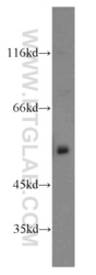
- Experimental details
- mouse brain tissue were subjected to SDS PAGE followed by western blot with 14306-1-AP(WASL antibody) at dilution of 1:500
- Sample type
- tissue
Supportive validation
- Submitted by
- Proteintech Group (provider)
- Main image

- Experimental details
- Immunofluorescent analysis of HepG2 cells, using WASL antibody 14306-1-AP at 1:25 dilution and Rhodamine-labeled goat anti-rabbit IgG (red). Blue pseudocolor = DAPI (fluorescent DNA dye).
- Sample type
- cell line

Supportive validation
- Submitted by
- Proteintech Group (provider)
- Main image
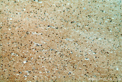
- Experimental details
- Immunohistochemical of paraffin-embedded human brain using 14306-1-AP(WASL antibody) at dilution of 1:50 (under 10x lens)
- Sample type
- tissue
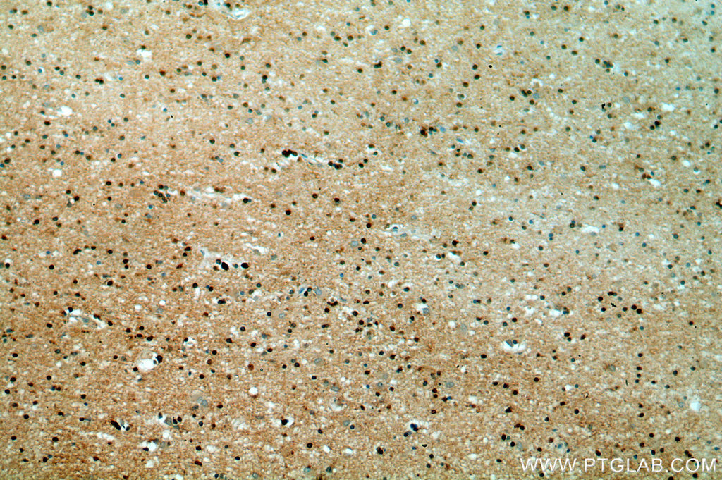
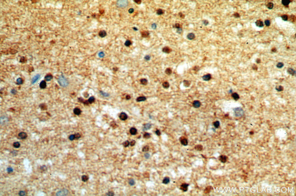
- Submitted by
- Proteintech Group (provider)
- Main image

- Experimental details
- Immunohistochemical of paraffin-embedded human brain using 14306-1-AP(WASL antibody) at dilution of 1:50 (under 40x lens)
- Sample type
- tissue