Explore
 Validate
 Learn
Learn Western blot
 Immunoprecipitation
Antibody data
- Antibody Data
- Antigen structure
- References [2]
- Comments [0]
- Validations
- Western blot [4]
Submit
Validation data
Reference
Comment
Report error
- Product number
- PA3-813 - Provider product page

- Provider
- Invitrogen Antibodies
- Product name
- RAR gamma-1 Polyclonal Antibody
- Antibody type
- Polyclonal
- Antigen
- Synthetic peptide
- Reactivity
- Human, Mouse, Rat
- Host
- Rabbit
- Isotype
- IgG
- Vial size
- 100 µL
- Concentration
- Conc. Not Determined
- Storage
- -20° C, Avoid Freeze/Thaw Cycles
Submitted references Pharmacologic retinoid signaling and physiologic retinoic acid receptor signaling inhibit basal cell carcinoma tumorigenesis.
The expression of retinoic acid receptor alpha is increased in the granule cells of the dentate gyrus in schizophrenia.
So PL, Fujimoto MA, Epstein EH Jr
Molecular cancer therapeutics 2008 May;7(5):1275-84
Molecular cancer therapeutics 2008 May;7(5):1275-84
The expression of retinoic acid receptor alpha is increased in the granule cells of the dentate gyrus in schizophrenia.
Rioux L, Arnold SE
Psychiatry research 2005 Jan 30;133(1):13-21
Psychiatry research 2005 Jan 30;133(1):13-21
No comments: Submit comment
Supportive validation
- Submitted by
- Invitrogen Antibodies (provider)
- Main image

- Experimental details
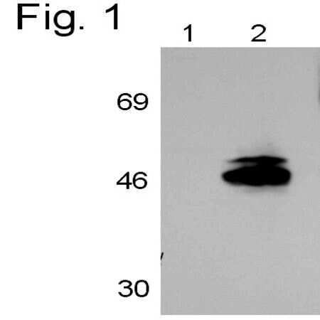
- WB detection of RAR gamma 1 from human HEK293 cells. Lane 1 shows PA3-813 incubated with the neutralizing peptide (PEP-228), lane 2 shows RAR detection by PA3-813.
- Submitted by
- Invitrogen Antibodies (provider)
- Main image

- Experimental details
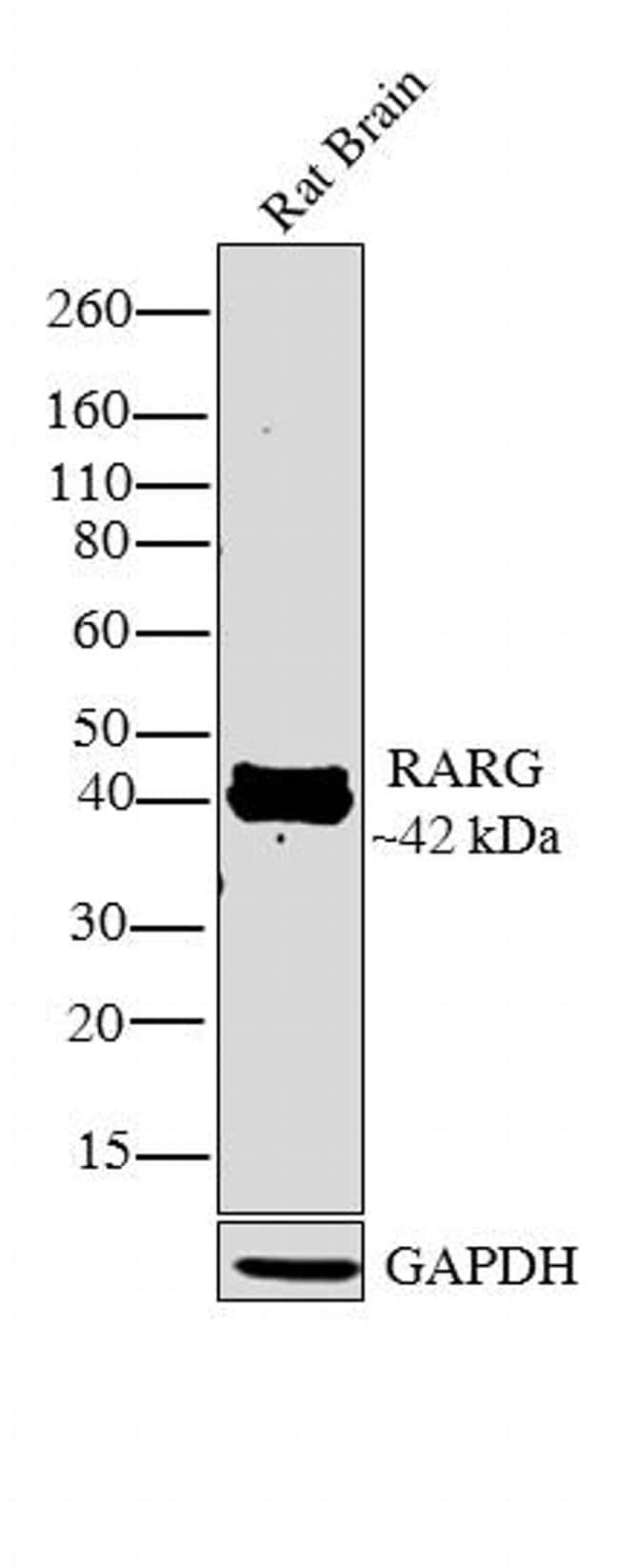
- Western blot analysis was performed on tissue extract (30 µg lysate) of Rat Brain (Lane 1). The blot was probed with Anti-RARG Rabbit Polyclonal Antibody (Product # PA3-813, 1:250 dilution) and detected by chemiluminescence using Goat anti-Rabbit IgG (H+L) Superclonal™ Secondary Antibody, HRP conjugate (Product # A27036, 0.25 µg/mL, 1:4000 dilution). A 42 kDa band corresponding to RARG was observed in the tissue tested. Known quantity of protein samples were electrophoresed using Novex® NuPAGE® 4-12 % Bis-Tris gel (Product # NP0322BOX), XCell SureLock™ Electrophoresis System (Product # EI0002) and Novex® Sharp Pre-Stained Protein Standard (Product # LC5800). Resolved proteins were then transferred onto a nitrocellulose membrane with iBlot® 2 Dry Blotting System (Product # IB21001). The membrane was probed with the relevant primary and secondary Antibody following blocking with 5% skimmed milk. Chemiluminescent detection was performed using Pierce™ ECL Western Blotting Substrate (Product # 32106).

- Submitted by
- Invitrogen Antibodies (provider)
- Main image

- Experimental details
- Western blot analysis was performed on tissue extract (30 µg lysate) of Rat Brain (Lane 1). The blot was probed with Anti-RARG Rabbit Polyclonal Antibody (Product # PA3-813, 1:250 dilution) and detected by chemiluminescence using Goat anti-Rabbit IgG (H+L) Superclonal™ Secondary Antibody, HRP conjugate (Product # A27036, 0.25 µg/mL, 1:4000 dilution). A 42 kDa band corresponding to RARG was observed in the tissue tested. Known quantity of protein samples were electrophoresed using Novex® NuPAGE® 4-12 % Bis-Tris gel (Product # NP0322BOX), XCell SureLock™ Electrophoresis System (Product # EI0002) and Novex® Sharp Pre-Stained Protein Standard (Product # LC5800). Resolved proteins were then transferred onto a nitrocellulose membrane with iBlot® 2 Dry Blotting System (Product # IB21001). The membrane was probed with the relevant primary and secondary Antibody following blocking with 5% skimmed milk. Chemiluminescent detection was performed using Pierce™ ECL Western Blotting Substrate (Product # 32106).
- Submitted by
- Invitrogen Antibodies (provider)
- Main image

- Experimental details
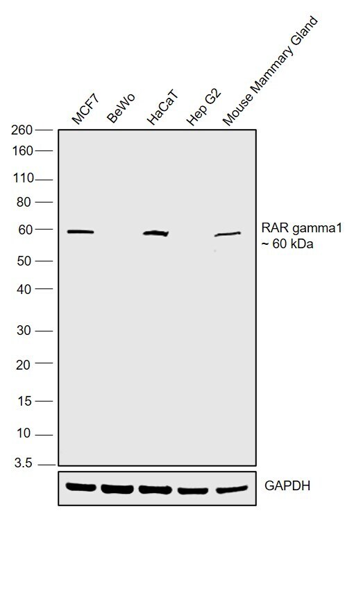
- Western blot was performed using Anti-RAR gamma-1 Polyclonal Antibody (Product # PA3-813) and a 60 kDa band corresponding to RAR gamma-1 was observed in MCF-7, HaCaT and Mouse Mammary Gland but not in BeWo and HepG2. Nuclear enriched extracts (50 µg lysate) of MCF7 (Lane 1), BeWo (Lane 2), HaCaT (Lane 3), Hep G2 (Lane 4) and tissue extract (50 µg lysate) of Mouse Mammary gland (Lane 5) were electrophoresed using NuPAGE™ 10% Bis-Tris Protein Gel (Product # NP0301BOX). Resolved proteins were then transferred onto a Nitrocellulose membrane (Product # IB23001) by iBlot® 2 Dry Blotting System (Product # IB21001). The blot was probed with the primary antibody (1:500) and detected by chemiluminescence with Goat anti-Rabbit IgG (H+L) Superclonal™ Recombinant Secondary Antibody, HRP (Product # A27036, 1:4000) using the iBright FL 1000 (Product # A32752). Chemiluminescent detection was performed using Novex® ECL Reagent Kit (Product # WP20005).