 Explore
 Validate
 Learn
Learn Western blot
Antibody data
- Antibody Data
- Antigen structure
- References [3]
- Comments [0]
- Validations
- Western blot [7]
- Immunocytochemistry [2]
- Immunohistochemistry [6]
- Other assay [2]
Submit
Validation data
Reference
Comment
Report error
- Product number
- PA5-22072 - Provider product page

- Provider
- Invitrogen Antibodies
- Product name
- Villin Polyclonal Antibody
- Antibody type
- Polyclonal
- Antigen
- Recombinant protein fragment
- Reactivity
- Human, Mouse, Rat
- Host
- Rabbit
- Isotype
- IgG
- Vial size
- 100 µL
- Concentration
- 0.85 mg/mL
- Storage
- Store at 4°C short term. For long term storage, store at -20°C, avoiding freeze/thaw cycles.
Submitted references A new murine esophageal organoid culture method and organoid-based model of esophageal squamous cell neoplasia.
Establishment of three novel cell lines derived from African American patients with colorectal carcinoma: A unique tool for assessing racial health disparity.
Loss of Cystic Fibrosis Transmembrane Regulator Impairs Intestinal Oxalate Secretion.
Zheng B, Ko KP, Fang X, Wang X, Zhang J, Jun S, Kim BJ, Luo W, Kim MJ, Jung YS, Cervantes CL, Park JI
iScience 2021 Dec 17;24(12):103440
iScience 2021 Dec 17;24(12):103440
Establishment of three novel cell lines derived from African American patients with colorectal carcinoma: A unique tool for assessing racial health disparity.
Paredes J, Ji P, Lacomb JF, Shroyer KR, Martello LA, Williams JL
International journal of oncology 2018 Oct;53(4):1516-1528
International journal of oncology 2018 Oct;53(4):1516-1528
Loss of Cystic Fibrosis Transmembrane Regulator Impairs Intestinal Oxalate Secretion.
Knauf F, Thomson RB, Heneghan JF, Jiang Z, Adebamiro A, Thomson CL, Barone C, Asplin JR, Egan ME, Alper SL, Aronson PS
Journal of the American Society of Nephrology : JASN 2017 Jan;28(1):242-249
Journal of the American Society of Nephrology : JASN 2017 Jan;28(1):242-249
No comments: Submit comment
Supportive validation
- Submitted by
- Invitrogen Antibodies (provider)
- Main image
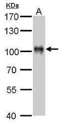
- Experimental details
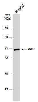
- Western blot analysis of Villin using 50 µg mouse kidney lysate. Samples were loaded onto a 7.5% SDS-PAGE gel and probed with a Villin polyclonal antibody (Product # PA5-22072) at a dilution of 1:1000.
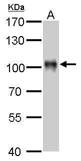
- Submitted by
- Invitrogen Antibodies (provider)
- Main image
- Experimental details
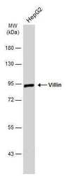
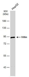
- Western blot analysis of Villin using 30 µg of HepG2 lysate. Samples were loaded onto a 7.5% SDS-PAGE gel and probed with a Villin polyclonal antibody (Product # PA5-22072) at a dilution of 1:5000.
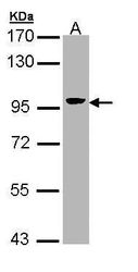
- Submitted by
- Invitrogen Antibodies (provider)
- Main image
- Experimental details
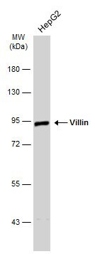
- Western Blot analysis of Villin was performed by separating 30 µg of Whole cell extracts by 7.5% SDS-PAGE. Proteins were transferred to a membrane and probed with a Villin Polyclonal Antibody (Product # PA5-22072) at a dilution of 1:2000. The HRP-conjugated anti-rabbit IgG antibody was used to detect the primary antibody.
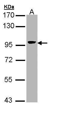
- Submitted by
- Invitrogen Antibodies (provider)
- Main image
- Experimental details
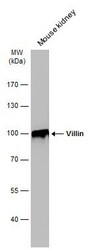
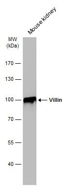
- Western Blot analysis of Villin was performed by separating 50 µg of Mouse tissue extracts by 7.5% SDS-PAGE. Proteins were transferred to a membrane and probed with a Villin Polyclonal Antibody (Product # PA5-22072) at a dilution of 1:1000. The HRP-conjugated anti-rabbit IgG antibody was used to detect the primary antibody.
- Submitted by
- Invitrogen Antibodies (provider)
- Main image

- Experimental details
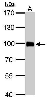
- Villin Polyclonal Antibody detects VIL1 protein by western blot analysis. A. 50 µg rat kidney lysate/extract.7.5% SDS-PAGE. Villin Polyclonal Antibody (Product # PA5-22072) dilution: 1:2,000. The HRP-conjugated anti-rabbit IgG antibody was used to detect the primary antibody.
- Submitted by
- Invitrogen Antibodies (provider)
- Main image
- Experimental details
- Western Blot analysis of Villin was performed by separating 30 µg of Whole cell extracts by 7.5% SDS-PAGE. Proteins were transferred to a membrane and probed with a Villin Polyclonal Antibody (Product # PA5-22072) at a dilution of 1:2000. The HRP-conjugated anti-rabbit IgG antibody was used to detect the primary antibody.
- Submitted by
- Invitrogen Antibodies (provider)
- Main image
- Experimental details
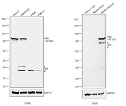
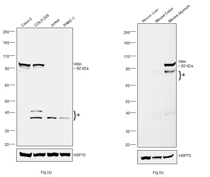
- Western blot was performed using Anti-Villin Polyclonal Antibody (Product # PA5-22072) and a 92kDa band corresponding to Villin was observed along with uncharacterized bands (*) across cell lines and tissues tested. Whole cell extracts (30 µg lysate) of Caco-2 (Lane 1), COLO 205 (Lane 2), Jurkat (Lane 3) and PANC-1 (Lane 4) as seen in Fig (a).Tissue extracts of Mouse Liver (Lane 1), Mouse Colon (Lane 2) and Mouse Jejunum (Lane 3) as seen in Fig (b) were electrophoresed using NuPAGE™ 4-12% Bis-Tris Protein Gel (Product # NP0321BOX). Relative expression of Villin was observed high in Caco-2 and COLO 205 and low to negative in Jurkat and PANC-1 as seen in Fig (a), similarly expression of Villin was high in Mouse Jejunum and low to negative in Mouse Colon and Mouse Liver as seen in Fig (b). Resolved proteins were then transferred onto a Nitrocellulose membrane (Product # IB23001) by iBlot® 2 Dry Blotting System (Product # IB21001). The blot was probed with the primary antibody (1:1000 dilution) and detected by chemiluminescence with Goat anti-Rabbit IgG (H+L) Superclonal™ Recombinant Secondary Antibody, HRP (Product # A27036, 1:4000 dilution) using the iBright FL 1000 (Product # A32752). Chemiluminescent detection was performed using Novex® ECL Chemiluminescent Substrate Reagent Kit (Product # WP20005).
Supportive validation
- Submitted by
- Invitrogen Antibodies (provider)
- Main image
- Experimental details
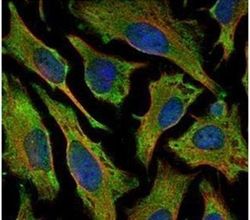
- Immunofluorescent analysis of Villin in methanol-fixed HeLa cells using a Villin polyclonal antibody (Product # PA5-22072) (Green) at a 1:500 dilution. Alpha-tubulin filaments were labeled with Product # PA5-29281 (Red) at a 1:2000.
- Submitted by
- Invitrogen Antibodies (provider)
- Main image

- Experimental details
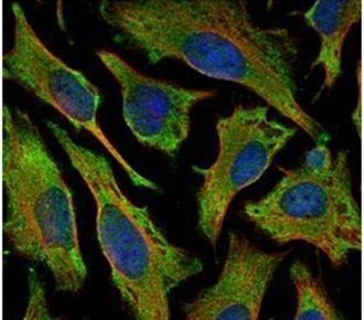
- Confocal immunofluorescence analysis (Olympus FV10i) of methanol-fixed HeLa, using Villin antibody (Product # PA5-22072) (Green) at 1:500 dilution. Alpha-tubulin filaments were labeled with (Product # MA1-25054) (Red) at 1:2,000.
Supportive validation
- Submitted by
- Invitrogen Antibodies (provider)
- Main image
- Experimental details
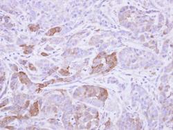
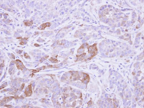
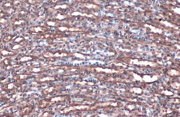
- Immunohistochemical analysis of paraffin-embedded A549 xenograft, using Villin (Product # PA5-22072) antibody at 1:500 dilution. Antigen Retrieval: EDTA based buffer, pH 8.0, 15 min.
- Submitted by
- Invitrogen Antibodies (provider)
- Main image

- Experimental details
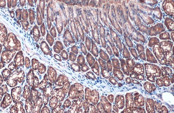
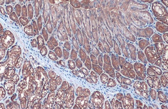
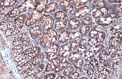
- Villin Polyclonal Antibody detects Villin protein at cytoplasm by immunohistochemical analysis. Sample: Paraffin-embedded mouse duodenum. Villin stained by Villin Polyclonal Antibody (Product # PA5-22072) diluted at 1:500. Antigen Retrieval: Citrate buffer, pH 6.0, 15 min.
- Submitted by
- Invitrogen Antibodies (provider)
- Main image

- Experimental details
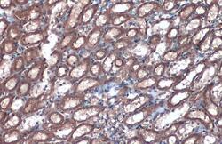
- Villin Polyclonal Antibody detects Villin protein at cytoplasm by immunohistochemical analysis. Sample: Paraffin-embedded mouse kidney. Villin stained by Villin Polyclonal Antibody (Product # PA5-22072) diluted at 1:500. Antigen Retrieval: Citrate buffer, pH 6.0, 15 min.
- Submitted by
- Invitrogen Antibodies (provider)
- Main image
- Experimental details
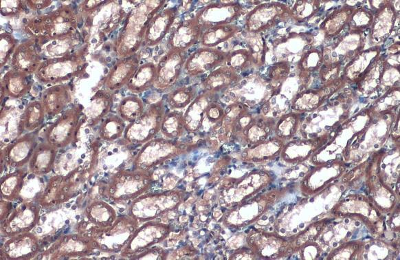
- Villin Polyclonal Antibody detects Villin protein at cytoplasm by immunohistochemical analysis. Sample: Paraffin-embedded rat colon. Villin stained by Villin Polyclonal Antibody (Product # PA5-22072) diluted at 1:500. Antigen Retrieval: Citrate buffer, pH 6.0, 15 min.

- Submitted by
- Invitrogen Antibodies (provider)
- Main image
- Experimental details
- Villin Polyclonal Antibody detects Villin protein at cytoplasm by immunohistochemical analysis. Sample: Paraffin-embedded rat kidney. Villin stained by Villin Polyclonal Antibody (Product # PA5-22072) diluted at 1:500. Antigen Retrieval: Citrate buffer, pH 6.0, 15 min.
Supportive validation
- Submitted by
- Invitrogen Antibodies (provider)
- Main image
- Experimental details
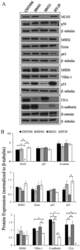
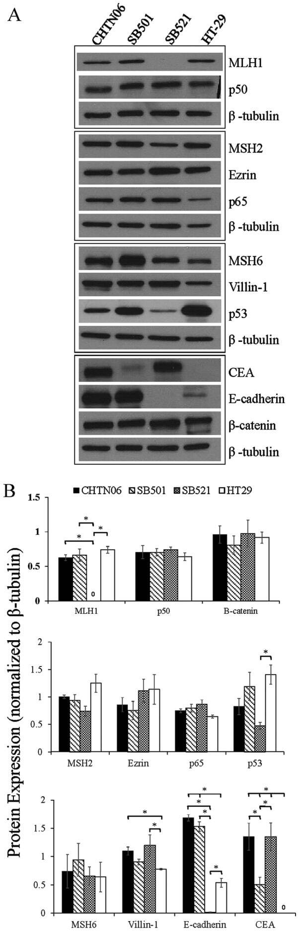
- Figure 3 The expression of proteins associated with colorectal carcinoma (CRC) tumorigenesis and metastasis was determined in the novel African American CRC lines by immunoblotting. (A) Qualitative analysis of CHTN06, SB501 and SB521 and HT-29, a Caucasian American CRC cell line, for protein expression of beta-catenin, p53, nuclear factor (NF)-kappaB (p50 and p65), villin-1, MSH2, MSH6, MLH1 and ezrin. (B) Semi-quantitative densitometry was performed by normalizing protein expression to the respective beta-tubulin loading control. Data were generated from three independent experiments. CEA, carcinoembryonic antigen.
- Submitted by
- Invitrogen Antibodies (provider)
- Main image
- Experimental details
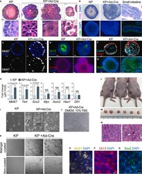
- Figure 5 Recapitulation of early ESCCs by Kras G12D :Trp53 KO model (A-I) Expression of esophageal epithelium neoplasia markers in EOs in E-MEOM. (A) H&E staining of KP+Ad-Cre EOs displayed loss of cell polarity, increased mitotic nuclei, nuclear polymorphism, and multiple nuclei. (B) ESCC-related neoplastic transformation. Possibility of EAC development was excluded by EAC marker immunostaining. PAS and Villin are not expressed in the KP+Ad-Cre organoids. (C-G) Increased expression of beta-catenin, Cd44, Ki67, and Sox2 in KP+Ad-Cre EOs. Immunofluorescence staining of Mki67 (C), Krt13 (D), beta-catenin (E), Cd44 (F), and Sox2 (G) for KP and KP+Ad-Cre EOs cultured in E-MEOM. (H) qRT-PCR for mRNA analysis of Ki67 , Tert , Sox2 , Myc , and Axin2 . Experiments were performed three times. (I-K) Neoplastic growth evaluation of Kras G12D :Trp53KO cell line in 2D culture model. (I) Bright-field images of KP+Ad-Cre versus KP cells cultured in a 24-well plate without Matrigel in EMEOM + Y-27632 (first 2 days). (J) Bright-field images of KP+Ad-Cre cells cultured in a 24-well plate in DMEM +10% FBS after three passages. (K) KP organoid cells and KP+Cre organoid cells were seeded on the Matrigel-coated or non-coated 2D culture plate. Cells were incubated for 2 weeks with medium change every 3 days, and differential interference contrast (DIC) images were taken by LSM-800. (L-Q) In vivo tumorigenicity of Kras G12D :Trp53KO EO cells. Kras G12D :Trp53KO EO cells (5 x 10 6 ) were subcutaneous
