 Explore
 Validate
 Learn
Learn Western blot
 Immunohistochemistry
Antibody data
- Antibody Data
- Antigen structure
- References [1]
- Comments [0]
- Validations
- Western blot [1]
- Immunocytochemistry [1]
- Immunohistochemistry [6]
Submit
Validation data
Reference
Comment
Report error
- Product number
- HPA006885 - Provider product page

- Provider
- Atlas Antibodies
- Proper citation
- Atlas Antibodies Cat#HPA006885, RRID:AB_1080564
- Product name
- Anti-VIL1
- Antibody type
- Polyclonal
- Reactivity
- Human
- Host
- Rabbit
- Conjugate
- Unconjugated
- Antigen sequence
NGPESTRMERLRGMTLAKEIRDQERGGRTYVGVVD
GENELASPKLMEVMNHVLGKRRELKAAVPDTVVEP
ALKAALKLYHVSDSEGNLVVREVATRPLTQDLLSH
E- Isotype
- IgG
- Vial size
- 100 µl
- Storage
- Store at +4°C for short term storage. Long time storage is recommended at -20°C.
Submitted references Scalable In Situ Hybridization on Tissue Arrays for Validation of Novel Cancer and Tissue-Specific Biomarkers
Kiflemariam S, Andersson S, Asplund A, Pontén F, Sjöblom T, Srivastava R
PLoS ONE 2012 March;7(3)
PLoS ONE 2012 March;7(3)
No comments: Submit comment
Supportive validation
- Submitted by
- Atlas Antibodies (provider)
- Enhanced method
- Independent antibody validation
- Main image
- Experimental details
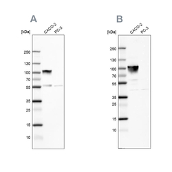
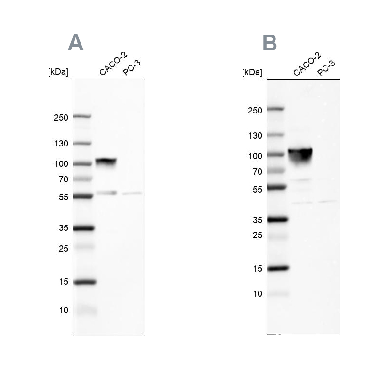
- Western blot analysis using Anti-VIL1 antibody HPA006885 (A) shows similar pattern to independent antibody HPA006884 (B).
Supportive validation
- Submitted by
- Atlas Antibodies (provider)
- Main image
- Experimental details
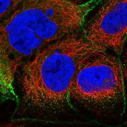
- Immunofluorescent staining of human cell line CACO-2 shows localization to plasma membrane.
- Sample type
- HUMAN

Enhanced validation
Supportive validation
- Submitted by
- Atlas Antibodies (provider)
- Enhanced method
- Independent antibody validation
- Main image
- Experimental details
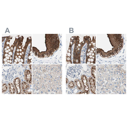
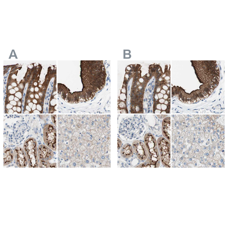
- Immunohistochemical staining of human colon, gallbladder, kidney and liver using Anti-VIL1 antibody HPA006885 (A) shows similar protein distribution across tissues to independent antibody HPA006884 (B).
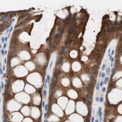
Supportive validation
- Submitted by
- Atlas Antibodies (provider)
- Main image

- Experimental details
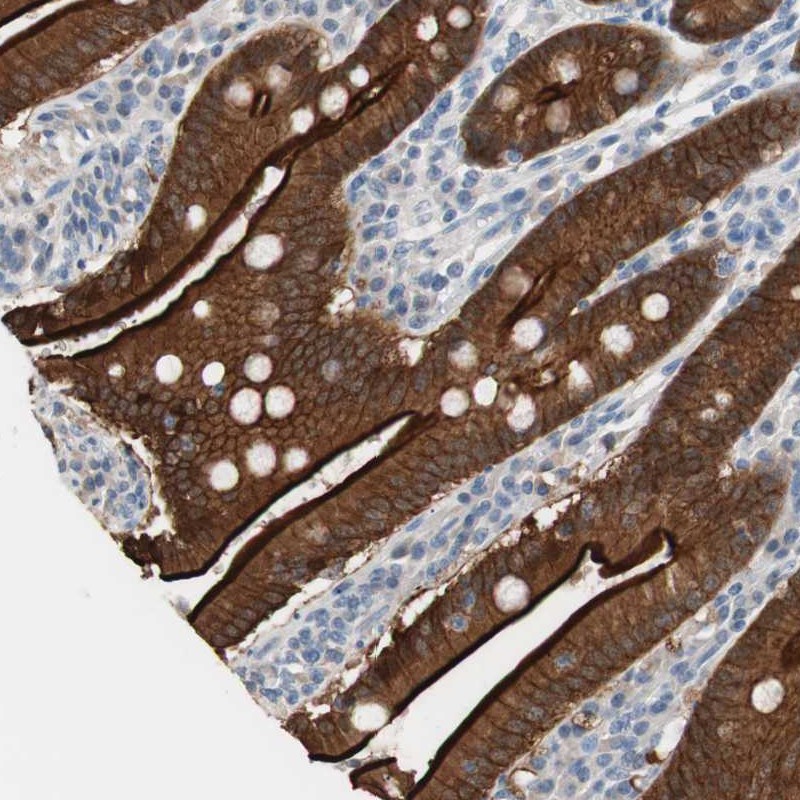
- Immunohistochemical staining of human duodenum shows strong luminal membranous and cytoplasmic positivity in glandular cells.
- Submitted by
- Atlas Antibodies (provider)
- Main image
- Experimental details
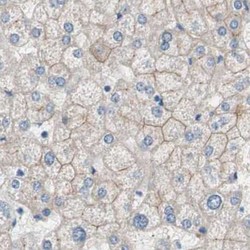
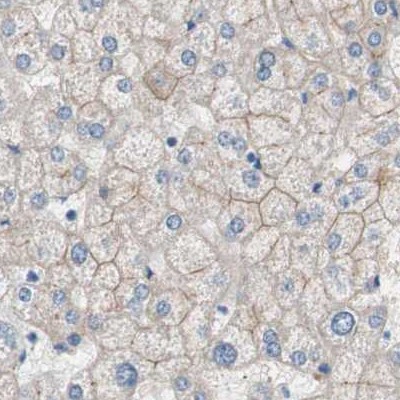
- Immunohistochemical staining of human liver using Anti-VIL1 antibody HPA006885.
- Sample type
- HUMAN
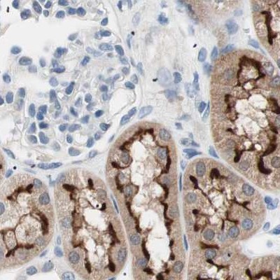
- Submitted by
- Atlas Antibodies (provider)
- Main image

- Experimental details
- Immunohistochemical staining of human kidney using Anti-VIL1 antibody HPA006885.
- Sample type
- HUMAN
- Submitted by
- Atlas Antibodies (provider)
- Main image
- Experimental details
- Immunohistochemical staining of human colon using Anti-VIL1 antibody HPA006885.
- Sample type
- HUMAN

- Submitted by
- Atlas Antibodies (provider)
- Main image

- Experimental details
- Immunohistochemical staining of human gallbladder using Anti-VIL1 antibody HPA006885.
- Sample type
- HUMAN
