Explore
 Validate
 Learn
Learn Western blot
 Immunohistochemistry
Antibody data
- Antibody Data
- Antigen structure
- References [1]
- Comments [0]
- Validations
- Western blot [4]
- Immunocytochemistry [1]
- Immunohistochemistry [13]
Submit
Validation data
Reference
Comment
Report error
- Product number
- HPA008057 - Provider product page

- Provider
- Atlas Antibodies
- Proper citation
- Atlas Antibodies Cat#HPA008057, RRID:AB_1851833
- Product name
- Anti-INA
- Antibody type
- Polyclonal
- Reactivity
- Human, Mouse, Rat
- Host
- Rabbit
- Conjugate
- Unconjugated
- Antigen sequence
ALDIEIAAYRKLLEGEETRFSTSGLSISGLNPLPN
PSYLLPPRILSATTSKVSSTGLSLKKEEEEEEASK
VASKKTSQIGESFEEILEETVISTKKTEKSNIEET
TISSQ- Isotype
- IgG
- Vial size
- 100 µl
- Storage
- Store at +4°C for short term storage. Long time storage is recommended at -20°C.
Submitted references Tissue Profiling of the Mammalian Central Nervous System Using Human Antibody-based Proteomics
Mulder J, Bjorling E, Jonasson K, Wernerus H, Hober S, Hokfelt T, Uhlen M
Molecular & Cellular Proteomics 2009 July;8(7):1612-1622
Molecular & Cellular Proteomics 2009 July;8(7):1612-1622
No comments: Submit comment
Supportive validation
- Submitted by
- Atlas Antibodies (provider)
- Main image
- Experimental details
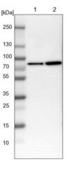
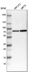
- Lane 1: NIH-3T3 cell lysate (Mouse embryonic fibroblast cells)Lane 2: NBT-II cell lysate (Rat Wistar bladder tumour cells)

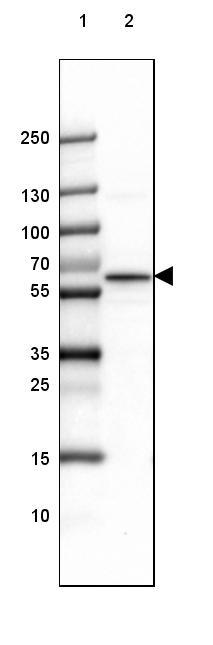
- Submitted by
- Atlas Antibodies (provider)
- Main image

- Experimental details
- Lane 1: Marker [kDa] 250, 130, 100, 70, 55, 35, 25, 15, 10Lane 2: Mouse Cerebral Cortex tissue
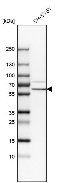
- Submitted by
- Atlas Antibodies (provider)
- Main image

- Experimental details
- Western blot analysis in human cell line SH-SY5Y.
- Submitted by
- Atlas Antibodies (provider)
- Main image
- Experimental details
- Western blot analysis in mouse cell line NIH-3T3 and rat cell line NBT-II.

Supportive validation
- Submitted by
- Atlas Antibodies (provider)
- Main image
- Experimental details
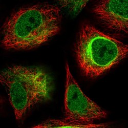
- Immunofluorescent staining of human cell line A549 shows localization to nucleoplasm, nuclear membrane & intermediate filaments.
- Sample type
- HUMAN

Enhanced validation
Supportive validation
- Submitted by
- Atlas Antibodies (provider)
- Enhanced method
- Orthogonal validation
- Main image
- Experimental details
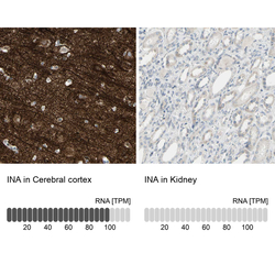
- Immunohistochemistry analysis in human cerebral cortex and kidney tissues using HPA008057 antibody. Corresponding INA RNA-seq data are presented for the same tissues.
- Sample type
- HUMAN

Supportive validation
- Submitted by
- Atlas Antibodies (provider)
- Main image
- Experimental details
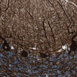
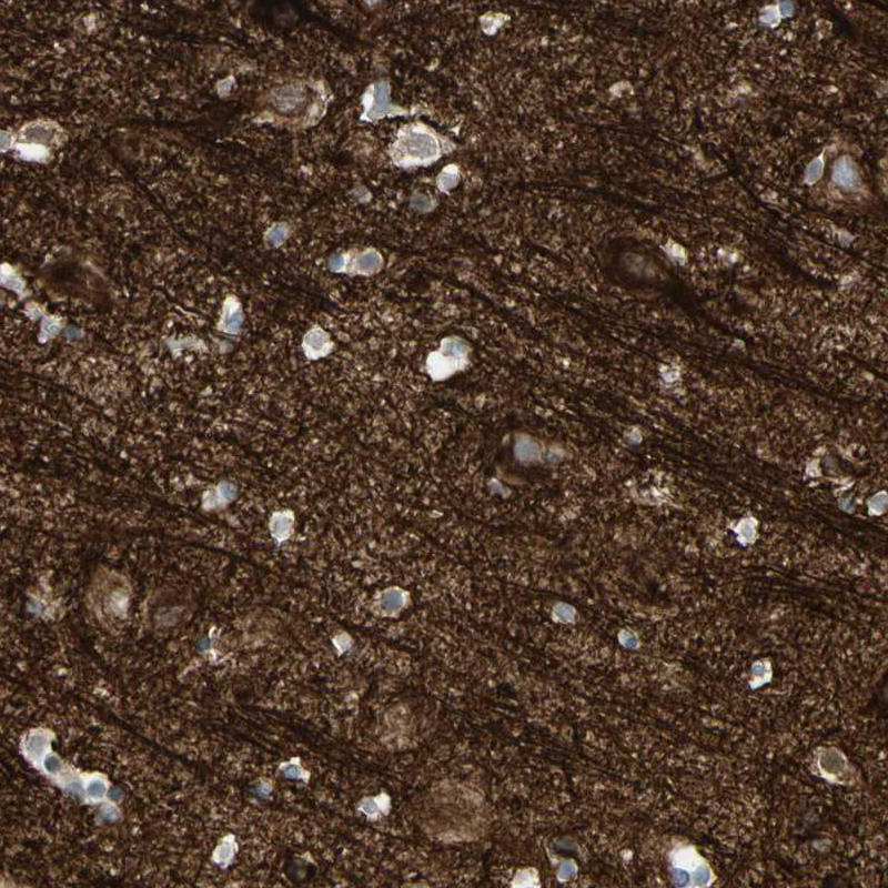
- Immunohistochemical staining of human cerebellum shows strong cytoplasmic positivity in purkinje cells and neuropil.
- Submitted by
- Atlas Antibodies (provider)
- Main image

- Experimental details
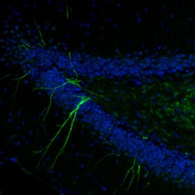
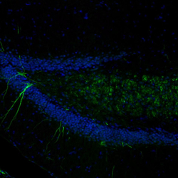
- Immunofluorescence staining of mouse dentate gyrus shows selective immunoreactivity in a subset of granular cells and their dendrites.
- Submitted by
- Atlas Antibodies (provider)
- Main image
- Experimental details
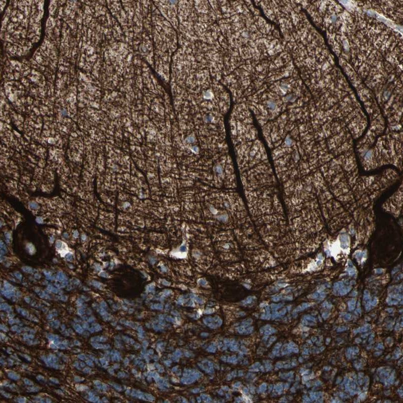
- Immunohistochemical staining of human hippocampus shows distinct positivity in neuronal processes.

- Submitted by
- Atlas Antibodies (provider)
- Main image

- Experimental details
- Immunofluorescence staining of mouse olfactory bulb shows strong positivity in neuronal processes and some cell bodies in the external plexiform layer.
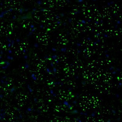
- Submitted by
- Atlas Antibodies (provider)
- Main image
- Experimental details
- Immunofluorescence staining of mouse dorsal tenia tecta shows selective immunoreactivity in a subset of neurons.
- Submitted by
- Atlas Antibodies (provider)
- Main image

- Experimental details
- Immunofluorescence staining of mouse medulla shows distinct staining of nerve bundles.
- Submitted by
- Atlas Antibodies (provider)
- Main image
- Experimental details
- Immunohistochemical staining oh human hippocampus shows strong positivity in neuropil.

- Submitted by
- Atlas Antibodies (provider)
- Main image
- Experimental details
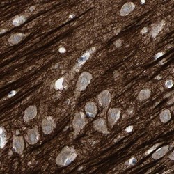
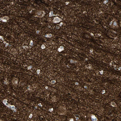
- Immunohistochemical staining of human cerebellum shows positivity in purkinje cells.
- Sample type
- HUMAN

- Submitted by
- Atlas Antibodies (provider)
- Main image
- Experimental details
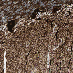
- Immunohistochemical staining of human cerebral cortex shows positivity in neuropil.
- Sample type
- HUMAN
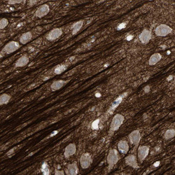
- Submitted by
- Atlas Antibodies (provider)
- Main image
- Experimental details
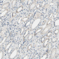
- Immunohistochemical staining of human kidney shows very weak cytoplasmic positivity.
- Sample type
- HUMAN

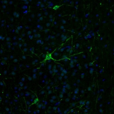
- Submitted by
- Atlas Antibodies (provider)
- Main image
- Experimental details
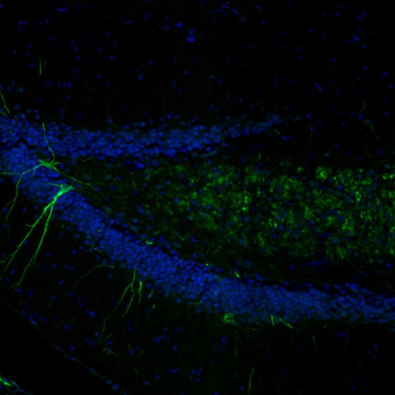
- Immunofluorescence staining of mouse brain shows moderate positivity in of in the hippocampus.
- Sample type
- MOUSE
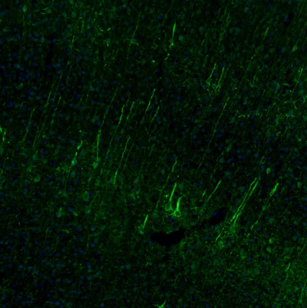
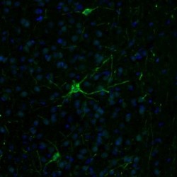
- Submitted by
- Atlas Antibodies (provider)
- Main image

- Experimental details
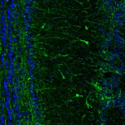
- Immunofluorescence staining of mouse brain shows positivity in of in the cerebral cortex.
- Sample type
- MOUSE