Explore
 Validate
 Learn
Learn Western blot
Antibody data
- Antibody Data
- Antigen structure
- References [7]
- Comments [0]
- Validations
- Western blot [2]
- Immunocytochemistry [1]
- Immunohistochemistry [1]
- Flow cytometry [1]
- Other assay [2]
Submit
Validation data
Reference
Comment
Report error
- Product number
- PA5-16681 - Provider product page

- Provider
- Invitrogen Antibodies
- Product name
- Calretinin Polyclonal Antibody
- Antibody type
- Polyclonal
- Antigen
- Recombinant full-length protein
- Reactivity
- Human, Mouse
- Host
- Rabbit
- Isotype
- IgG
- Vial size
- 500 µL
- Storage
- -20° C, Avoid Freeze/Thaw Cycles
Submitted references The Interaction between Reactive Peritoneal Mesothelial Cells and Tumor Cells via Extracellular Vesicles Facilitates Colorectal Cancer Dissemination.
Transcriptome analysis of fetal rat testis following intrauterine exposure to the azole fungicides triticonazole and flusilazole reveals subtle changes despite adverse endocrine effects.
Preparation of Gelatin and Gelatin/Hyaluronic Acid Cryogel Scaffolds for the 3D Culture of Mesothelial Cells and Mesothelium Tissue Regeneration.
Amacrine cells coupled to ganglion cells via gap junctions are highly vulnerable in glaucomatous mouse retinas.
Mesothelial cells facilitate cancer stem‑like properties in spheroids of ovarian cancer cells.
Granulosa Cell Tumors: Novel Predictors of Recurrence in Early-stage Patients.
Targeting neuronal gap junctions in mouse retina offers neuroprotection in glaucoma.
Serratì S, Porcelli L, Fragassi F, Garofoli M, Di Fonte R, Fucci L, Iacobazzi RM, Palazzo A, Margheri F, Cristiani G, Albano A, De Luca R, Altomare DF, Simone M, Azzariti A
Cancers 2021 May 20;13(10)
Cancers 2021 May 20;13(10)
Transcriptome analysis of fetal rat testis following intrauterine exposure to the azole fungicides triticonazole and flusilazole reveals subtle changes despite adverse endocrine effects.
Draskau MK, Lardenois A, Evrard B, Boberg J, Chalmel F, Svingen T
Chemosphere 2021 Feb;264(Pt 1):128468
Chemosphere 2021 Feb;264(Pt 1):128468
Preparation of Gelatin and Gelatin/Hyaluronic Acid Cryogel Scaffolds for the 3D Culture of Mesothelial Cells and Mesothelium Tissue Regeneration.
Kao HH, Kuo CY, Chen KS, Chen JP
International journal of molecular sciences 2019 Sep 12;20(18)
International journal of molecular sciences 2019 Sep 12;20(18)
Amacrine cells coupled to ganglion cells via gap junctions are highly vulnerable in glaucomatous mouse retinas.
Akopian A, Kumar S, Ramakrishnan H, Viswanathan S, Bloomfield SA
The Journal of comparative neurology 2019 Jan 1;527(1):159-173
The Journal of comparative neurology 2019 Jan 1;527(1):159-173
Mesothelial cells facilitate cancer stem‑like properties in spheroids of ovarian cancer cells.
Shishido A, Mori S, Yokoyama Y, Hamada Y, Minami K, Qian Y, Wang J, Hirose H, Wu X, Kawaguchi N, Nagumo S, Matsuura N, Yamamoto H
Oncology reports 2018 Oct;40(4):2105-2114
Oncology reports 2018 Oct;40(4):2105-2114
Granulosa Cell Tumors: Novel Predictors of Recurrence in Early-stage Patients.
Sakr S, Abdulfatah E, Thomas S, Al-Wahab Z, Beydoun R, Morris R, Ali-Fehmi R, Bandyopadhyay S
International journal of gynecological pathology : official journal of the International Society of Gynecological Pathologists 2017 May;36(3):240-252
International journal of gynecological pathology : official journal of the International Society of Gynecological Pathologists 2017 May;36(3):240-252
Targeting neuronal gap junctions in mouse retina offers neuroprotection in glaucoma.
Akopian A, Kumar S, Ramakrishnan H, Roy K, Viswanathan S, Bloomfield SA
The Journal of clinical investigation 2017 Jun 30;127(7):2647-2661
The Journal of clinical investigation 2017 Jun 30;127(7):2647-2661
No comments: Submit comment
Supportive validation
- Submitted by
- Invitrogen Antibodies (provider)
- Main image

- Experimental details
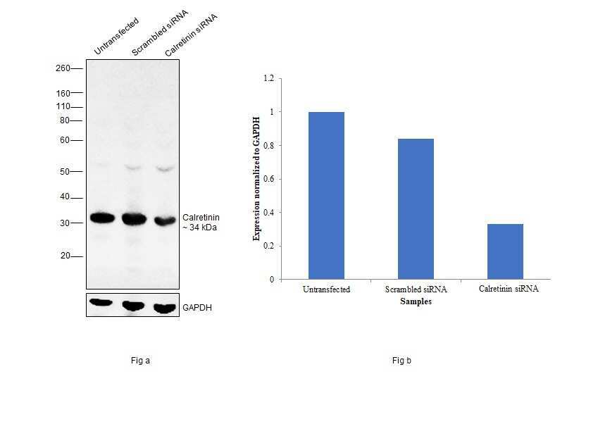
- Knockdown of Calretinin was achieved by transfecting HT-29 with Calretinin specific siRNAs (Silencer® select Product # s224821, s2325). Western blot analysis (Fig. a) was performed using membrane enriched extracts from the Calretinin knockdown cells (lane 3), non-specific scrambled siRNA transfected cells (lane 2) and untransfected cells (lane 1). The blot was probed with Calretinin Polyclonal Antibody (Product # PA5-16881, 1:25 dilution) and Goat anti-Rabbit IgG (H+L), Superclonal™ Recombinant Secondary Antibody, HRP (Product # A27036, 1:4000 dilution). Densitometric analysis of this western blot is shown in histogram (Fig. b). Decrease in signal upon siRNA mediated knock down confirms that antibody is specific to Calretinin.
- Submitted by
- Invitrogen Antibodies (provider)
- Main image
- Experimental details
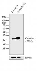
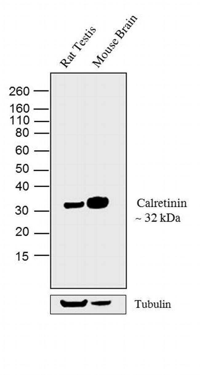
- Western blot analysis was performed on tissue extracts (30 µg lysate) of Rat Testis (Lane 1) and Mouse Brain (Lane 2).The blots were probed with Calretinin Rabbit polyclonal Antibody (Product # PA5-16681, 2 µg/mL) and detected by chemiluminescence using Goat anti-Rabbit IgG (H+L) Superclonal™ Secondary Antibody, HRP conjugate (Product # A27036, 0.4 µg/mL, 1:2500 dilution). A 32 kDa band corresponding to Calretinin was observed across the tissues tested. Known quantity of protein samples were electrophoresed using Novex® NuPAGE® 4-12 % Bis-Tris gel (Product # NP0322BOX), XCell SureLock™ Electrophoresis System (Product # EI0002) and Novex® Sharp Pre-Stained Protein Standard (Product # LC5800). Resolved proteins were then transferred onto a nitrocellulose membrane with iBlot® 2 Dry Blotting System (Product # IB21001). The membrane was probed with the relevant primary and secondary Antibody following blocking with 5% skimmed milk. Chemiluminescent detection was performed using Pierce™ ECL Western Blotting Substrate (Product # 32106).
Supportive validation
- Submitted by
- Invitrogen Antibodies (provider)
- Main image

- Experimental details
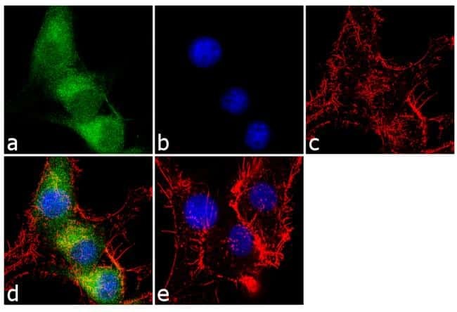
- Immunofluorescence analysis of Calretinin was performed using 70% confluent log phase U-87 MG cells. The cells were fixed with 4% paraformaldehyde for 10 minutes, permeabilized with 0.1% Triton™ X-100 for 10 minutes, and blocked with 1% BSA for 1 hour at room temperature. The cells were labeled with Calretinin Rabbit Polyclonal Antibody (Product # PA5-16681) at 2µg/mL in 0.1% BSA and incubated for 3 hours at room temperature and then labeled with Goat anti-Rabbit IgG (H+L) Superclonal™ Secondary Antibody, Alexa Fluor® 488 conjugate (Product # A27034) at a dilution of 1:2000 for 45 minutes at room temperature (Panel a: green). Nuclei (Panel b: blue) were stained with SlowFade® Gold Antifade Mountant with DAPI (Product # S36938). F-actin (Panel c: red) was stained with Alexa Fluor® 555 Rhodamine Phalloidin (Product # R415, 1:300). Panel d represents the merged image showing cytoplasmic localization. Panel e shows the no primary antibody control. The images were captured at 60X magnification.
Supportive validation
- Submitted by
- Invitrogen Antibodies (provider)
- Main image
- Experimental details
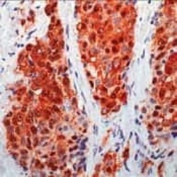
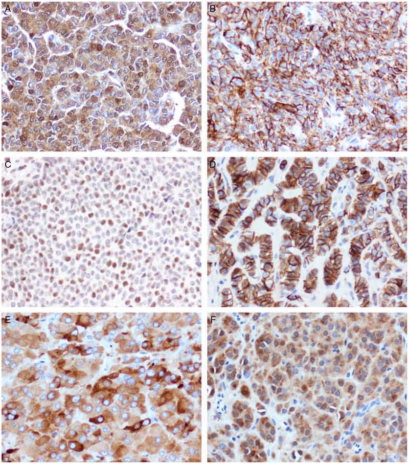
- Formalin-fixed, paraffin-embedded human mesothelioma stained with Calretinin using peroxidase-conjugate and AEC chromogen. Note cytoplasmic and nuclear staining of tumor cells.

Supportive validation
- Submitted by
- Invitrogen Antibodies (provider)
- Main image
- Experimental details
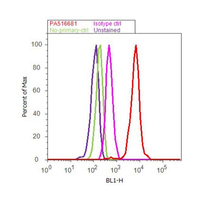
- Flow cytometry analysis of Calretinin was done on U-87 MG cells. Cells were fixed with 70% ethanol for 10 minutes, permeabilized with 0.25% Triton™ X-100 for 20 minutes, and blocked with 5% BSA for 30 minutes at room temperature. Cells were labeled with Calretinin Rabbit Polyclonal Antibody (PA5-16681, red histogram) or with rabbit isotype control (pink histogram) at 3-5 µg/million cells in 2.5% BSA. After incubation at room temperature for 2 hours, the cells were labeled with Alexa Fluor® 488 Goat Anti-Rabbit Secondary Antibody (A11008) at a dilution of 1:400 for 30 minutes at room temperature. The representative 10,000 cells were acquired and analyzed for each sample using an Attune® Acoustic Focusing Cytometer. The purple histogram represents unstained control cells and the green histogram represents no-primary-antibody control..
Supportive validation
- Submitted by
- Invitrogen Antibodies (provider)
- Main image

- Experimental details
- NULL
- Submitted by
- Invitrogen Antibodies (provider)
- Main image
- Experimental details
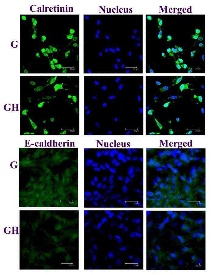
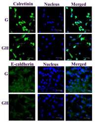
- Figure 7 The immunofluorescence (IF) staining of calretinin and E-cadherin of the mesothelial cells cultured in G and GH for seven days. The protein was stained green by a fluorescein isothiocyanate (FITC)-conjugated secondary antibody, while the nuclei were stained blue by Hoechst 33342. Bar = 30 mum.