Explore
 Validate
 Learn
Learn Western blot
Antibody data
- Antibody Data
- Antigen structure
- References [1]
- Comments [0]
- Validations
- Western blot [1]
- Immunocytochemistry [1]
- Immunohistochemistry [1]
- Other assay [1]
Submit
Validation data
Reference
Comment
Report error
- Product number
- PA5-51930 - Provider product page

- Provider
- Invitrogen Antibodies
- Product name
- LTBP2 Polyclonal Antibody
- Antibody type
- Polyclonal
- Antigen
- Recombinant full-length protein
- Description
- Immunogen sequence: ERSPNLRRSS AAGEGTLARA QPPAPQSPPA PQSPPAGTLS GLSQTHPSQQ HVGLSRTVRL HPTATASSQL SSNALPPGPG LEQRDGTQQA VPLEHPSSPW GLNLTEKIKK IKIVFTPTIC KQTCARGHCA NSCERG
- Concentration
- 0.3 mg/mL
Submitted references N6-Methyladenosine Regulates the Expression and Secretion of TGFβ1 to Affect the Epithelial-Mesenchymal Transition of Cancer Cells.
Li J, Chen F, Peng Y, Lv Z, Lin X, Chen Z, Wang H
Cells 2020 Jan 25;9(2)
Cells 2020 Jan 25;9(2)
No comments: Submit comment
Supportive validation
- Submitted by
- Invitrogen Antibodies (provider)
- Main image

- Experimental details
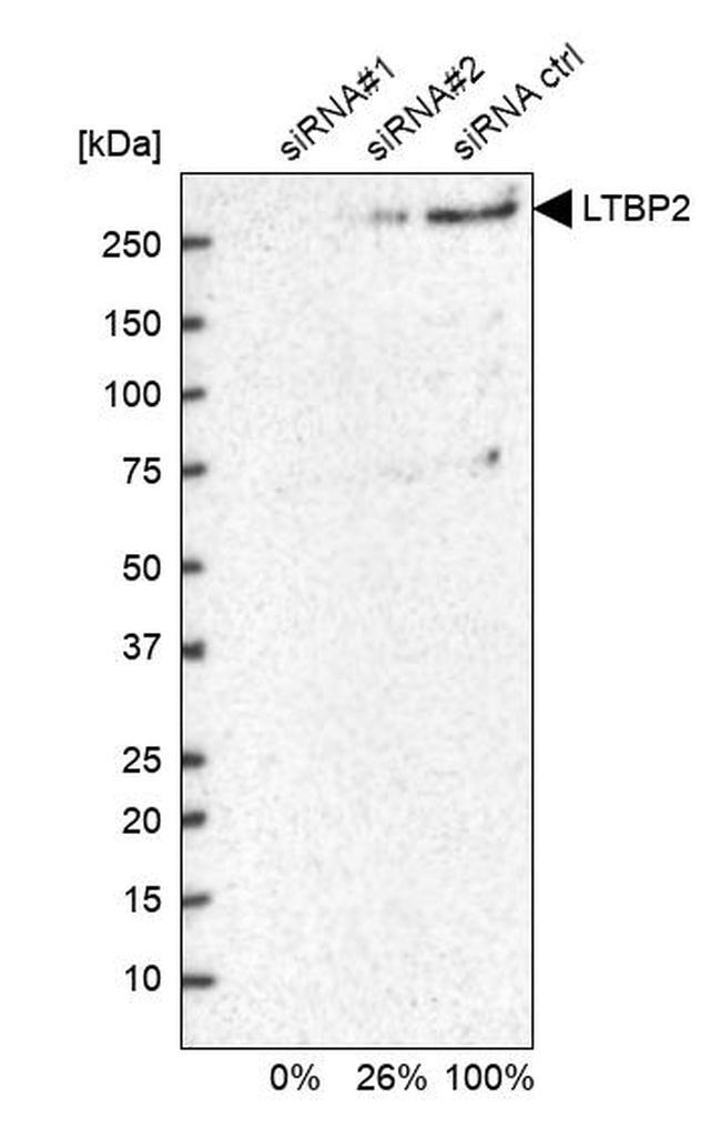
- Western blot analysis of LTBP2 in U2OS cells transfected with control siRNA, target specific siRNA probe #1 and #2, using a LTBP2 Polyclonal Antibody (Product # PA5-51930). Remaining relative intensity is presented.
Supportive validation
- Submitted by
- Invitrogen Antibodies (provider)
- Main image

- Experimental details
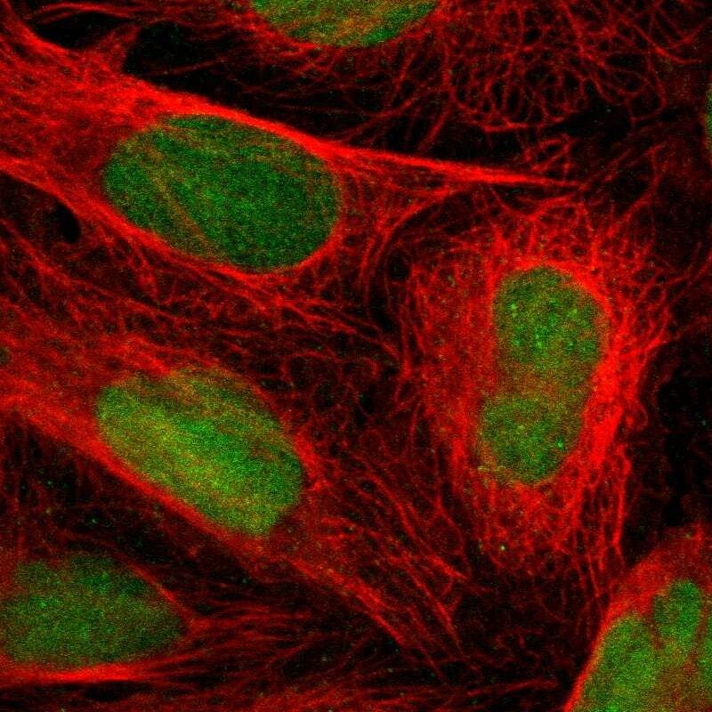
- Immunofluorescent staining of LTBP2 in human cell line U-2 OS using a LTBP2 Polyclonal Antibody (Product # PA5-51930) shows localization to nucleus.
Supportive validation
- Submitted by
- Invitrogen Antibodies (provider)
- Main image
- Experimental details
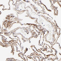
- Immunohistochemical staining of LTBP2 in human lung using a LTBP2 Polyclonal Antibody (Product # PA5-51930) shows strong positivity in extracellular matrix.

Supportive validation
- Submitted by
- Invitrogen Antibodies (provider)
- Main image

- Experimental details
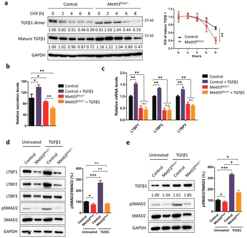
- Figure 5 Secretion of TGFbeta1 is modulated by METTL3. ( a ) Control and Mettl3 Mut/- HeLa cells were incubated with 100 µg/mL cycloheximide (CHX) for indicated times. Protein levels of mature TGFbeta1 and TGFbeta1 dimer were measured by Western blot (left). Band intensities were analyzed by ImageJ and are listed at the bottom of target bands. Mature TGFbeta1 levels were quantitatively analyzed (right); ( b ) Control and Mettl3 Mut/- HeLa cells were incubated with 10 ng/mL TGFbeta1 for 48 h. Secretion of TGFbeta1 in control and Mettl3 Mut/- HeLa cells was measured by ELISA kit. The relative secretion levels of TGFbeta1 were normalized to culture medium with or without the addition of TGFbeta1; ( c ) Control and Mettl3 Mut/- HeLa cells were incubated with 10 ng/mL TGFbeta1 for 48 h. The expression levels of LTBP1, LTBP2, and LTBP3 mRNA in control and Mettl3 Mut/- HeLa cells were measured by qRT-PCR; ( d ) Control and Mettl3 Mut/- HeLa cells were incubated with 10 ng/mL TGFbeta1 for 48 h. The expression levels of LTBP1, LTBP2, LTBP3, pSMAD2, and SMAD2 in control and Mettl3 Mut/- HeLa cells were measured by Western blot (left). Percentages of pSMAD2 to SMAD2 were analyzed (right); ( e ) Control and Mettl3 Mut/- HeLa cells were transiently overexpressed in LTBP1 for 24 h, then incubated with 10 ng/mL TGFbeta1 for 48 h. The expression levels of TGFbeta1, pSMAD2, and SMAD2 were measured by Western blot (left). Band intensities of TGFbeta1 were analyzed by ImageJ and are liste