 Explore
 Validate
 Learn
Learn Western blot
Antibody data
- Antibody Data
- Antigen structure
- References [0]
- Comments [0]
- Validations
- Western blot [4]
- Immunocytochemistry [1]
Submit
Validation data
Reference
Comment
Report error
- Product number
- PA5-78536 - Provider product page

- Provider
- Invitrogen Antibodies
- Product name
- PBK Polyclonal Antibody
- Antibody type
- Polyclonal
- Antigen
- Recombinant full-length protein
- Description
- Positive Control: 293T, A431, HeLa, HepG2, U87-MG, SK-N-SH, IMR32, SK-N-AS
- Concentration
- 1 mg/mL
No comments: Submit comment
Supportive validation
- Submitted by
- Invitrogen Antibodies (provider)
- Main image
- Experimental details
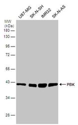
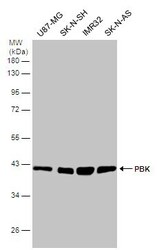
- Western blot analysis of PBK in whole cell lysate using 30 µg of protein. Samples were separated with 10% SDS-PAGE and incubated with PBK polyclonal antibody (Product # PA5-78536) using a dilution of 1:1000.
- Submitted by
- Invitrogen Antibodies (provider)
- Main image
- Experimental details
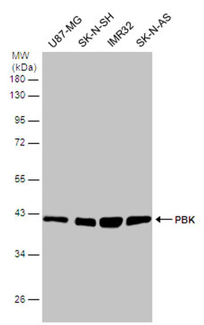
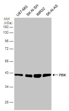
- Western blot analysis of PBK in whole cell lysate using 30 µg of protein. Samples were separated with 10% SDS-PAGE and incubated with PBK polyclonal antibody (Product # PA5-78536) using a dilution of 1:1000.
- Submitted by
- Invitrogen Antibodies (provider)
- Main image
- Experimental details
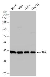
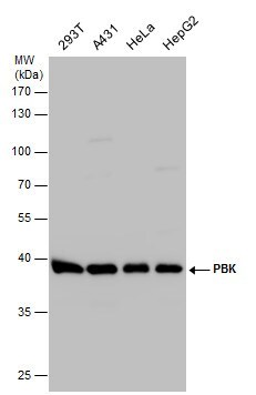
- Western Blot using PBK Polyclonal Antibody (Product # PA5-78536). Various whole cell extracts (30 µg) were separated by 10% SDS-PAGE, and the membrane was blotted with PBK Polyclonal Antibody (Product # PA5-78536) diluted at 1:1,000.
- Submitted by
- Invitrogen Antibodies (provider)
- Main image

- Experimental details
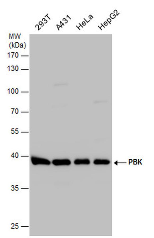
- PBK Polyclonal Antibody detects PBK protein by western blot analysis. Various whole cell extracts (30 µg) were separated by 10% SDS-PAGE, and the membrane was blotted with PBK Polyclonal Antibody (Product # PA5-78536) diluted at 1:1,000.
Supportive validation
- Submitted by
- Invitrogen Antibodies (provider)
- Main image
- Experimental details
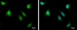
- PBK Polyclonal Antibody detects PBK protein at nucleus by immunofluorescent analysis. Sample: HeLa cells were fixed in 4% paraformaldehyde at RT for 15 min. Green: PBK protein stained by PBK Polyclonal Antibody (Product # PA5-78536) diluted at 1:500. Blue: Hoechst 33342 staining. Scale bar = 10 µm.
