Explore
 Validate
 Learn
Learn Western blot
 ELISA
ELISAAntibody data
- Antibody Data
- Antigen structure
- References [4]
- Comments [0]
- Validations
- Western blot [1]
- Immunocytochemistry [1]
- Flow cytometry [1]
- Blocking/Neutralizing [1]
Submit
Validation data
Reference
Comment
Report error
- Product number
- AF740 - Provider product page

- Provider
- R&D Systems
- Product name
- Mouse B7-1/CD80 Antibody
- Antibody type
- Polyclonal
- Description
- Antigen Affinity-purified. Detects mouse B7-1/CD80 in ELISAs and Western blots.
- Reactivity
- Mouse
- Host
- Goat
- Conjugate
- Unconjugated
- Antigen sequence
Q00609- Isotype
- IgG
- Vial size
- 100 ug
- Concentration
- LYOPH
- Storage
- Use a manual defrost freezer and avoid repeated freeze-thaw cycles. 12 months from date of receipt, -20 to -70 °C as supplied. 1 month, 2 to 8 °C under sterile conditions after reconstitution. 6 months, -20 to -70 °C under sterile conditions after reconstitution.
Submitted references Frontline Science: High fat diet and leptin promote tumor progression by inducing myeloid-derived suppressor cells.
Aging is associated with increased regulatory T-cell function.
Antigen-loaded ER microsomes from APC induce potent immune responses against viral infection.
Suppression of activation and induction of apoptosis in RAW264.7 cells by amniotic membrane extract.
Clements VK, Long T, Long R, Figley C, Smith DMC, Ostrand-Rosenberg S
Journal of leukocyte biology 2018 Mar;103(3):395-407
Journal of leukocyte biology 2018 Mar;103(3):395-407
Aging is associated with increased regulatory T-cell function.
Garg SK, Delaney C, Toubai T, Ghosh A, Reddy P, Banerjee R, Yung R
Aging cell 2014 Jun;13(3):441-8
Aging cell 2014 Jun;13(3):441-8
Antigen-loaded ER microsomes from APC induce potent immune responses against viral infection.
Sofra V, Mansour S, Liu M, Gao B, Primpidou E, Wang P, Li S
European journal of immunology 2009 Jan;39(1):85-95
European journal of immunology 2009 Jan;39(1):85-95
Suppression of activation and induction of apoptosis in RAW264.7 cells by amniotic membrane extract.
He H, Li W, Chen SY, Zhang S, Chen YT, Hayashida Y, Zhu YT, Tseng SC
Investigative ophthalmology & visual science 2008 Oct;49(10):4468-75
Investigative ophthalmology & visual science 2008 Oct;49(10):4468-75
No comments: Submit comment
Supportive validation
- Submitted by
- R&D Systems (provider)
- Main image
- Experimental details
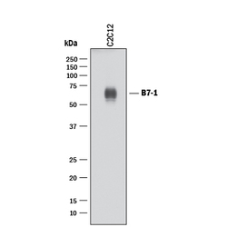
- Detection of Mouse B7-1/CD80 by Western Blot. Western blot shows lysates of C2C12 mouse myoblast cell line. PVDF membrane was probed with 1 µg/mL of Goat Anti-Mouse B7-1/CD80 Antigen Affinity-purified Polyclonal Antibody (Catalog # AF740) followed by HRP-conjugated Anti-Goat IgG Secondary Antibody (Catalog # HAF017). A specific band was detected for B7-1/CD80 at approximately 60 kDa (as indicated). This experiment was conducted under reducing conditions and using Immunoblot Buffer Group 1.

Supportive validation
- Submitted by
- R&D Systems (provider)
- Main image
- Experimental details
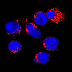
- B7-1/CD80 in Mouse Splenocytes. B7-1/CD80 was detected in immersion fixed mouse splenocytes using Goat Anti-Mouse B7-1/CD80 Antigen Affinity-purified Polyclonal Antibody (Catalog # AF740) at 15 µg/mL for 3 hours at room temperature. Cells were stained using the NorthernLights™ 557-conjugated Anti-Goat IgG Secondary Antibody (red; Catalog # NL001) and counterstained with DAPI (blue). Specific staining was localized to cytoplasm. View our protocol for Fluorescent ICC Staining of Non-adherent Cells.

Supportive validation
- Submitted by
- R&D Systems (provider)
- Main image

- Experimental details
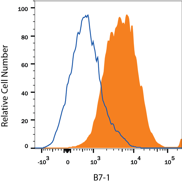
- Detection of B7-1/CD80 in Mouse Splenocytes by Flow Cytometry. Mouse splenocytes either treated with 200 ng/mL LPS (filled histogram) or unstimulated (open histogram) were stained with Goat Anti-Mouse B7-1/CD80 Antigen Affinity-purified Polyclonal Antibody (Catalog # AF740), followed by Phycoerythrin-conjugated Anti-Goat IgG Secondary Antibody (Catalog # F0107). View our protocol for Staining Membrane-associated Proteins.
Supportive validation
- Submitted by
- R&D Systems (provider)
- Main image

- Experimental details
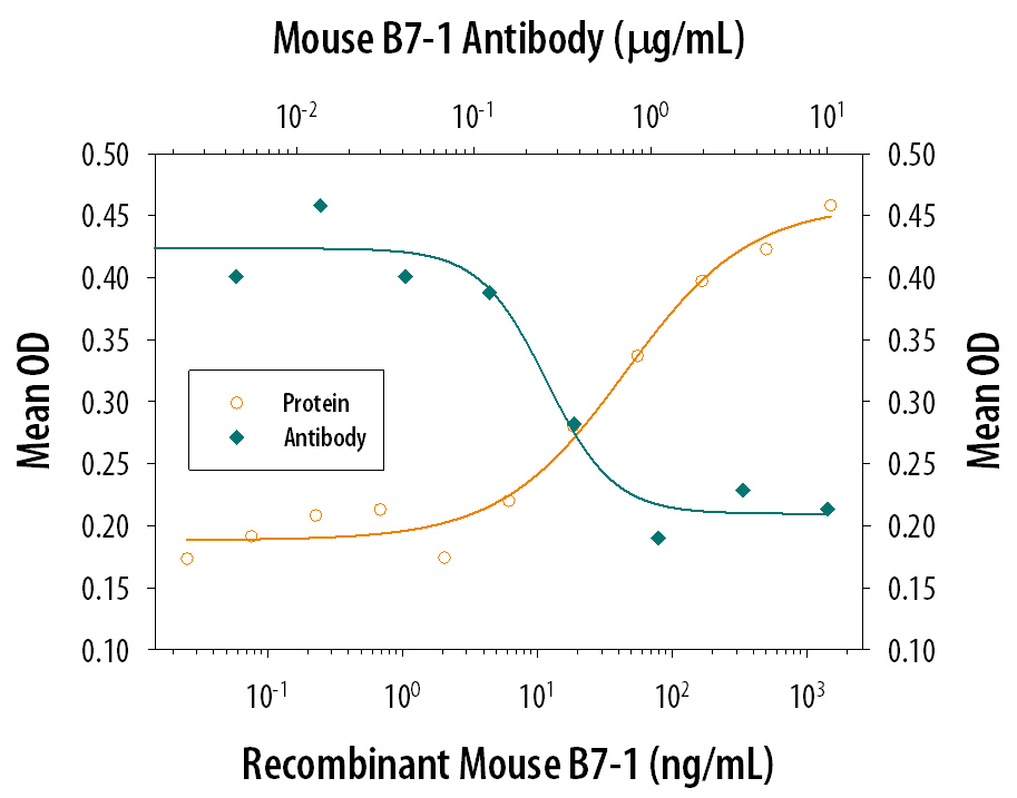
- IL-2 secretion Induced by B7-1/CD80 and Neutralization by Mouse B7-1/CD80 Antibody. Recombinant Mouse B7-1/CD80 Fc Chimera (Catalog # 740-B1) co-stimulates IL-2 secretion in the Jurkat human acute T cell leukemia cell line in the presence of PHA in a dose-dependent manner (orange line), as measured by the Human IL-2 Quantikine ELISA Kit (Catalog # D2050). IL-2 secretion elicited by Recombinant Mouse B7-1/CD80 Fc Chimera (0.1 µg/mL) and PHA (10 µg/mL) is neutralized (green line) by increasing concentrations of Goat Anti-Mouse B7-1/CD80 Antigen Affinity-purified Polyclonal Antibody (Catalog # AF740). The ND50 is typically 0.15-0.6 µg/mL.