Explore
 Validate
 Learn
Learn Western blot
Antibody data
- Antibody Data
- Antigen structure
- References [0]
- Comments [0]
- Validations
- Western blot [2]
- Immunohistochemistry [3]
Submit
Validation data
Reference
Comment
Report error
- Product number
- PA5-105919 - Provider product page

- Provider
- Invitrogen Antibodies
- Product name
- Phospho-TBK1 (Ser172) Polyclonal Antibody
- Antibody type
- Polyclonal
- Antigen
- Synthetic peptide
- Reactivity
- Human, Mouse, Rat
- Host
- Rabbit
- Isotype
- IgG
- Vial size
- 100 µL
- Concentration
- 1 mg/mL
- Storage
- -20°C
No comments: Submit comment
Supportive validation
- Submitted by
- Invitrogen Antibodies (provider)
- Main image
- Experimental details
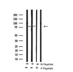
- Western blot analysis of Phospho-TBK1 (Ser172) in PMA treated HeLa cell lysate (absence (-) or presence (+) of non-phospho and phospho peptide). Samples were incubated with Phospho-TBK1 (Ser172) polyclonal antibody (Product # PA5-105919).

- Submitted by
- Invitrogen Antibodies (provider)
- Main image
- Experimental details
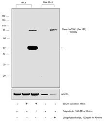
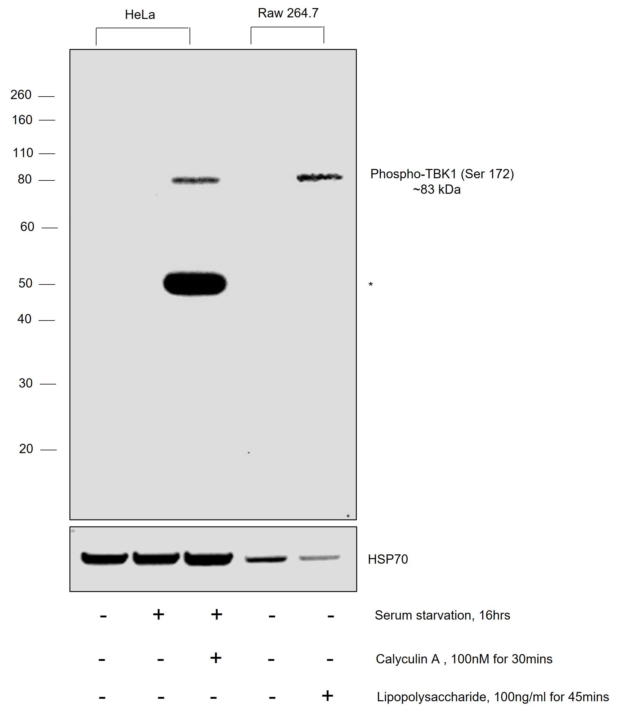
- Western blot was performed using Phospho-TBK1 (Ser172) Polyclonal Antibody (Product # PA5-105919) and a 83 kDa band corresponding to Phospho-TBK1 (Ser172) was observed across in serum starved HeLa cells upon treatment with Calyculin A and in RAW 264.7 cells upon treatment with Lipopolysaccharide. Whole cell extracts (30 µg lysate) of HeLa (Lane 1), HeLa (serum starved for 16hrs) (Lane 2), HeLa (serum starved for 16hrs and treated with 100 nM of Calyculin A for 30 mins) (Lane 3), RAW 264.7 (Lane 4), RAW 264.7 (treated with 100 ng/mL of Lipopolysaccharide for 45 mins) (Lane 5) were electrophoresed using NuPAGE™ 4-12% Bis-Tris Protein Gel (Product # NP0321BOX), 10 well. Resolved proteins were then transferred onto a nitrocellulose membrane (Product # IB23001) by iBlot® 2 Dry Blotting System (Product # IB21001). The blot was probed with the primary antibody (1:1000 dilution) and detected by chemiluminescence with Goat anti-Rabbit IgG (H+L) Superclonal™ Recombinant Secondary Antibody, HRP (Product # A27036, 1:20,000 dilution) using the iBright™ FL1500 Imaging System (Product # A44115). Chemiluminescent detection was performed using SuperSignal™ West Dura Extended Duration Substrate (Product # 34076).An uncharacterized band (*) at ~50 kDa was also induced in HeLa cells upon the same treatment.
Supportive validation
- Submitted by
- Invitrogen Antibodies (provider)
- Main image

- Experimental details
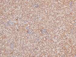
- Immunohistochemistry analysis of paraffin-embedded Phospho-TBK1 (Ser172) in human tonsil tissue sections. Antigen retrieval was performed using citrate buffer. Samples were blocked with blocking buffer (1.5 hr, 22°C), incubated with Phospho-TBK1 (Ser172) polyclonal antibody (Product # PA5-105919) using a dilution of 1:200 (1.5 hr, 22°C), followed by HRP conjugated goat anti-rabbit.

- Submitted by
- Invitrogen Antibodies (provider)
- Main image
- Experimental details
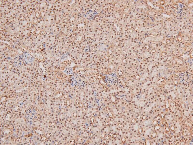
- Immunohistochemistry analysis of paraffin-embedded Phospho-TBK1 (Ser172) in mouse kidney tissue sections. Antigen retrieval was performed using citrate buffer. Samples were blocked with blocking buffer (1.5 hr, 22°C), incubated with Phospho-TBK1 (Ser172) polyclonal antibody (Product # PA5-105919) using a dilution of 1:200 (1.5 hr, 22°C), followed by HRP conjugated goat anti-rabbit.
- Submitted by
- Invitrogen Antibodies (provider)
- Main image

- Experimental details
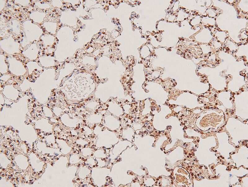
- Immunohistochemistry analysis of paraffin-embedded Phospho-TBK1 (Ser172) in rat lung tissue sections. Antigen retrieval was performed using citrate buffer. Samples were blocked with blocking buffer (1.5 hr, 22°C), incubated with Phospho-TBK1 (Ser172) polyclonal antibody (Product # PA5-105919) using a dilution of 1:200 (1.5 hr, 22°C), followed by HRP conjugated goat anti-rabbit.