 Explore
 Validate
 Learn
Learn Western blot
Antibody data
- Antibody Data
- Antigen structure
- References [0]
- Comments [0]
- Validations
- Western blot [3]
- Immunocytochemistry [3]
- Immunohistochemistry [5]
Submit
Validation data
Reference
Comment
Report error
- Product number
- PA5-52431 - Provider product page

- Provider
- Invitrogen Antibodies
- Product name
- WWTR1 Polyclonal Antibody
- Antibody type
- Polyclonal
- Antigen
- Recombinant full-length protein
- Description
- Immunogen sequence: MNPKPSSWRK KILPESFFKE PDSGSHSRQS STDSSGGHPG PRLAGGAQHV RSHSSPASLQ LGTGAGAAGS PAQQHAHLRQ QSYDVTDELP LPPGWEMTFT ATGQRYFLNH IEKITTWQDP RKAMNQPLNH MNLHPAVSST
- Concentration
- 0.1 mg/mL
No comments: Submit comment
Supportive validation
- Submitted by
- Invitrogen Antibodies (provider)
- Main image
- Experimental details
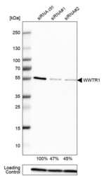
- Western blot analysis of WWTR1 in EFO-21 cells transfected with control siRNA, target specific siRNA probe #1 and #2, using a WWTR1 Polyclonal Antibody (Product # PA5-52431). Remaining relative intensity is presented. Loading control: Anti-PPIB.

- Submitted by
- Invitrogen Antibodies (provider)
- Main image
- Experimental details
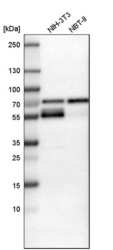
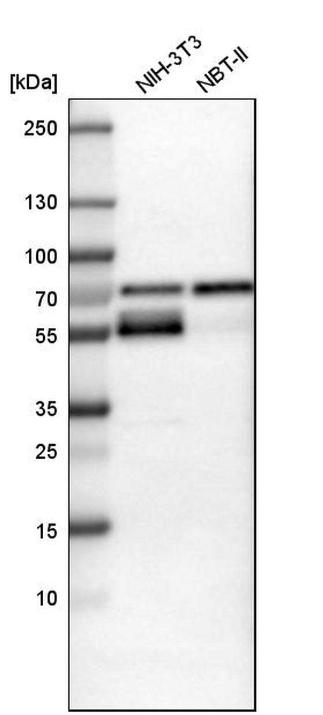
- Western blot analysis of WWTR1 in mouse cell line NIH-3T3 and rat cell line NBT-II using a WWTR1 Polyclonal Antibody (Product # PA5-52431).
- Submitted by
- Invitrogen Antibodies (provider)
- Main image
- Experimental details
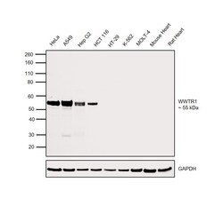
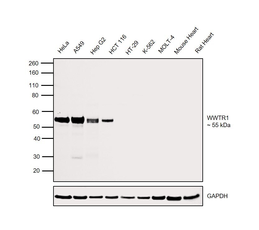
- Western blot was performed using Anti-WWTR1 Polyclonal Antibody (Product # PA5-52431) and a 55kDa band corresponding to WWTR1 was observed across cell lines tested except HT-29, K-562 and MOLT-4. This product does not show any reactivity to Mouse and Rat tissues. Whole cell extracts (30 µg lysate) of HeLa (Lane 1), A549 (Lane 2), Hep G2 (Lane 3), HCT 116 (Lane 4), HT-29 (Lane 5), K-562 (Lane 6), MOLT-4 (Lane 7), Mouse Heart (Lane 8), Rat Heart (Lane 9) were electrophoresed using NuPAGE™ 4-12% Bis-Tris Protein Gel (Product # NP0322BOX). Resolved proteins were then transferred onto a Nitrocellulose membrane (Product # IB23001) by iBlot® 2 Dry Blotting System (Product # IB21001). The blot was probed with the primary antibody (0.4 µg/mL) and detected by chemiluminescence with Goat anti-Rabbit IgG (H+L) Superclonal™ Recombinant Secondary Antibody, HRP (Product # A27036, 1:4000) using the iBright FL 1000 (Product # A32752). Chemiluminescent detection was performed using Novex® ECL Chemiluminescent Substrate Reagent Kit (Product # WP20005).
Supportive validation
- Submitted by
- Invitrogen Antibodies (provider)
- Main image
- Experimental details
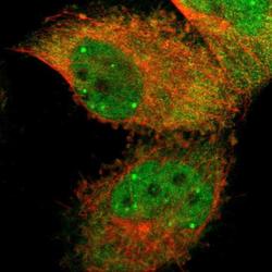
- Immunofluorescent staining of WWTR1 in human cell line U-251 MG shows positivity in cytoplasm & nucleus but excluded from the nucleoli. Samples were probed using a WWTR1 Polyclonal Antibody (Product # PA5-52431).

- Submitted by
- Invitrogen Antibodies (provider)
- Main image
- Experimental details
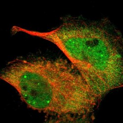
- Immunofluorescent staining of WWTR1 in human cell line U-251 MG using a WWTR1 Polyclonal Antibody (Product # PA5-52431) shows localization to nucleoplasm, nuclear bodies and cytosol.

- Submitted by
- Invitrogen Antibodies (provider)
- Main image
- Experimental details
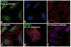
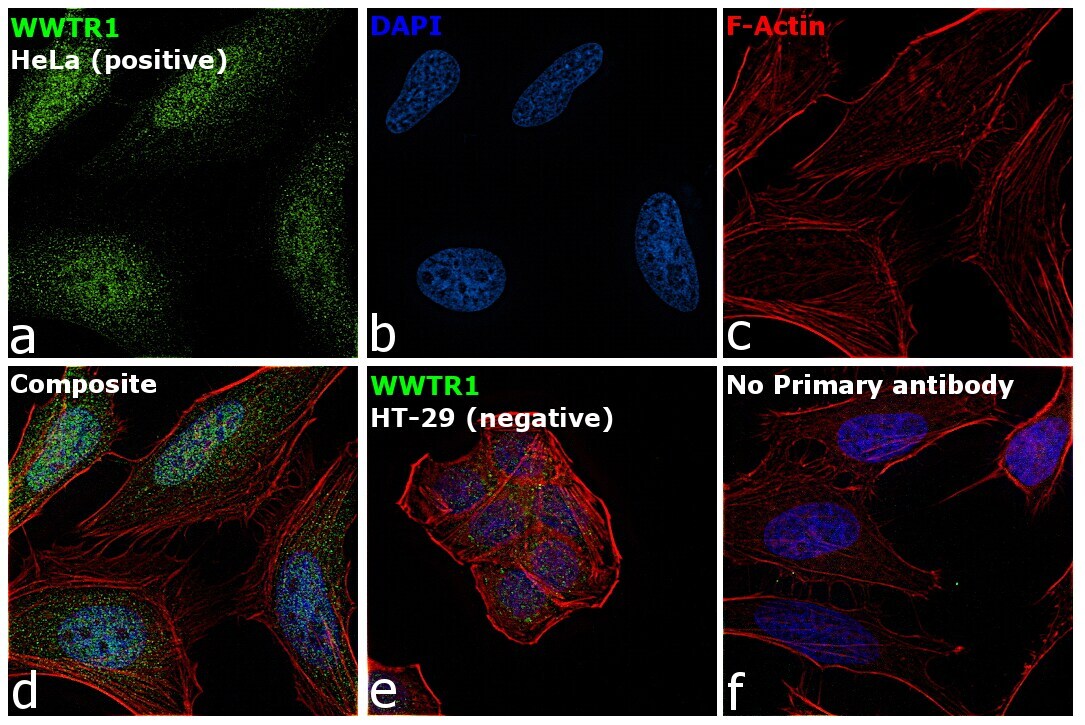
- Immunofluorescence analysis of WWTR1 was performed using 70% confluent log phase HeLa and HT-29 cells. The cells were fixed with 4% paraformaldehyde for 10 minutes, permeabilized with 0.1% Triton™ X-100 for 15 minutes, and blocked with 2% BSA for 45 minutes at room temperature. The cells were labeled with WWTR1 Polyclonal Antibody (Product # PA5-52431) at 1 µg/mL in 0.1% BSA, incubated at 4 degree celsius overnight and then labeled with Donkey anti-Rabbit IgG (H+L) Highly Cross-Adsorbed Secondary Antibody, Alexa Fluor Plus 488 (Product # A32790), (1:2000), for 45 minutes at room temperature (Panel a: Green). Nuclei (Panel b:Blue) were stained with ProLong™ Diamond Antifade Mountant with DAPI (Product # P36962). F-actin (Panel c: Red) was stained with Rhodamine Phalloidin (Product # R415, 1:300). Panel d represents the merged image of HeLa cells, which is a positive model for WWTR1 expression showing a nuclear and cytoplasmic localization. Panel e represents the merged image of HT-29 cells, that are null for WWTR1 protein expression. Panel f represents control cells with no primary antibody to assess background. The images were captured at 60X magnification.
Supportive validation
- Submitted by
- Invitrogen Antibodies (provider)
- Main image

- Experimental details
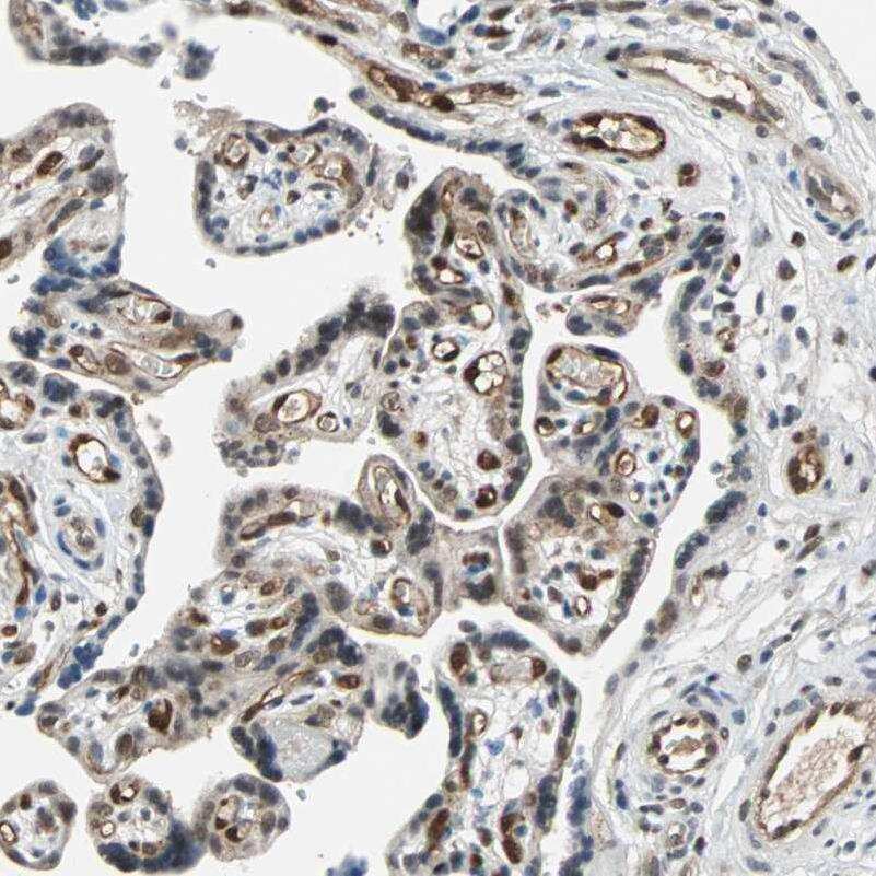
- Immunohistochemical staining of WWTR1 in human placenta using a WWTR1 Polyclonal Antibody (Product # PA5-52431) shows moderate to strong nuclear positivity in endothelial cells.
- Submitted by
- Invitrogen Antibodies (provider)
- Main image
- Experimental details
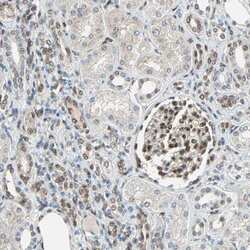
- Immunohistochemical staining of WWTR1 in human kidney using a WWTR1 Polyclonal Antibody (Product # PA5-52431) shows moderate nuclear positivity in glomerular cells and a subset of cells in distal tubules.

- Submitted by
- Invitrogen Antibodies (provider)
- Main image
- Experimental details
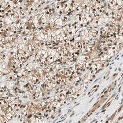
- Immunohistochemical staining of WWTR1 in human renal cancer using a WWTR1 Polyclonal Antibody (Product # PA5-52431) shows strong nuclear positivity in tumor cells.

- Submitted by
- Invitrogen Antibodies (provider)
- Main image
- Experimental details
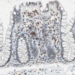
- Immunohistochemical staining of WWTR1 in human colon using WWTR1 Polyclonal Antibody (Product # PA5-52431) shows moderate to strong nuclear positivity in a subset of lymphoid cells.
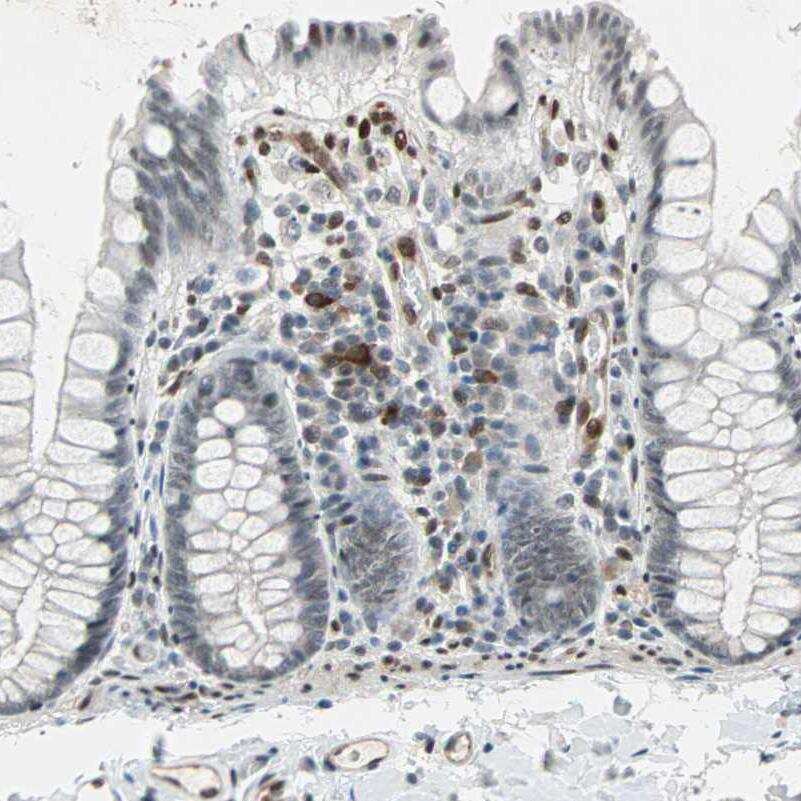
- Submitted by
- Invitrogen Antibodies (provider)
- Main image
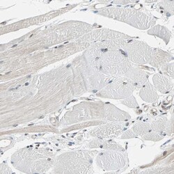
- Experimental details
- Immunohistochemical staining of WWTR1 in human skeletal muscle using WWTR1 Polyclonal Antibody (Product # PA5-52431) shows weak positivity in myocytes.
