Explore
 Validate
 Learn
LearnBAF1916
antibody from R&D Systems
Targeting: TP63
EEC3, KET, NBP, OFC8, p51, p53CP, p63, p73H, p73L, SHFM4, TP53CP, TP53L, TP73L
 Western blot
 Immunocytochemistry
Antibody data
- Antibody Data
- Antigen structure
- References [0]
- Comments [0]
- Validations
- Immunocytochemistry [1]
- Immunohistochemistry [1]
Submit
Validation data
Reference
Comment
Report error
- Product number
- BAF1916 - Provider product page

- Provider
- R&D Systems
- Product name
- Human p63/TP73L Biotinylated Antibody
- Antibody type
- Polyclonal
- Description
- Antigen Affinity-purified. Detects p63/TP73L in Western blots.
- Reactivity
- Human
- Host
- Goat
- Conjugate
- Biotin
- Antigen sequence
Q9H3D4- Isotype
- IgG
- Vial size
- 50 ug
- Storage
- Use a manual defrost freezer and avoid repeated freeze-thaw cycles. 12 months from date of receipt, -20 to -70 °C as supplied. 1 month, 2 to 8 °C under sterile conditions after reconstitution. 6 months, -20 to -70 °C under sterile conditions after reconstitution.
No comments: Submit comment
Supportive validation
- Submitted by
- R&D Systems (provider)
- Main image
- Experimental details
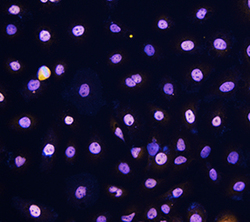
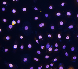
- p63/TP73L in SCC-25 Human Cell Line. p63/TP73L was detected in immersion fixed SCC-25 human tongue carcinoma cell line using Goat Anti-Human p63/TP73L Biotinylated Antigen Affinity-purified Polyclonal Antibody (Catalog # BAF1916) at 10 µg/mL for 3 hours at room temperature. Cells were stained using the NorthernLights™ 557-conjugated Streptavidin (yellow; Catalog # NL999) and counterstained with DAPI (blue). Specific staining was localized to nuclei. View our protocol for Fluorescent ICC Staining of Cells on Coverslips.
Supportive validation
- Submitted by
- R&D Systems (provider)
- Main image
- Experimental details
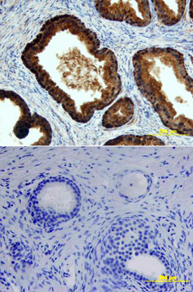
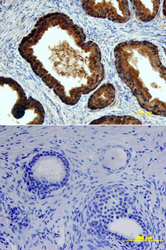
- p63/TP73L in Human Prostate. p63/TP73L was detected in immersion fixed paraffin-embedded sections of human prostate array using Goat Anti-Human p63/TP73L Biotinylated Antigen Affinity-purified Polyclonal Antibody (Catalog # BAF1916) at 15 µg/mL overnight at 4 °C. Tissue was stained using the Anti-Goat HRP-DAB Cell & Tissue Staining Kit (brown; Catalog # CTS008) and counterstained with hematoxylin (blue). Lower panel shows a lack of labeling if primary antibodies are omitted and tissue is stained only with secondary antibody followed by incubation with detection reagents. View our protocol for Chromogenic IHC Staining of Paraffin-embedded Tissue Sections.