Explore
 Validate
 Learn
Learn Western blot
Antibody data
- Antibody Data
- Antigen structure
- References [0]
- Comments [0]
- Validations
- Western blot [3]
- Immunohistochemistry [1]
Submit
Validation data
Reference
Comment
Report error
- Product number
- PA5-111751 - Provider product page

- Provider
- Invitrogen Antibodies
- Product name
- LRRC8A (extracellular) Polyclonal Antibody
- Antibody type
- Polyclonal
- Antigen
- Synthetic peptide
- Reactivity
- Human, Mouse, Rat
- Host
- Rabbit
- Isotype
- IgG
- Vial size
- 50 µL
- Concentration
- 0.8 mg/mL
- Storage
- -20°C
No comments: Submit comment
Supportive validation
- Submitted by
- Invitrogen Antibodies (provider)
- Main image
- Experimental details
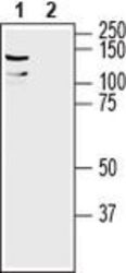
- Western Blot analysis of LRRC8A was performed in mouse brain lysate. Lane 1: LRRC8A (extracellular) Antibody (Product # PA5-111751) at a dilution of 1:200. Lane 2: LRRC8A (extracellular) Antibody preincubated with the negative control antigen.

- Submitted by
- Invitrogen Antibodies (provider)
- Main image

- Experimental details
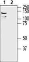
- Western Blot analysis of LRRC8A was performed in rat lung membranes. Lane 1: LRRC8A (extracellular) Antibody (Product # PA5-111751) at a dilution of 1:200. Lane 2: LRRC8A (extracellular) Antibody preincubated with the negative control antigen.

- Submitted by
- Invitrogen Antibodies (provider)
- Main image
- Experimental details
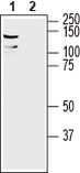
- Western Blot analysis of LRRC8A was performed in mouse brain lysate. Lane 1: LRRC8A (extracellular) Antibody (Product # PA5-111751) at a dilution of 1:200. Lane 2: LRRC8A (extracellular) Antibody preincubated with the negative control antigen.
Supportive validation
- Submitted by
- Invitrogen Antibodies (provider)
- Main image
- Experimental details
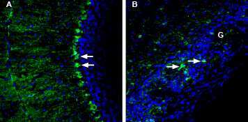
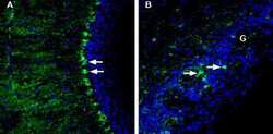
- Immunohistochemistry analysis of LRRC8A in perfusion-fixed, frozen mouse cerebellum and olfactory bulb tissue sections using LRRC8A (extracellular) Antibody (Product # PA5-111751) at a dilution of 1:200, followed by goat-anti-rabbit-AlexaFluor-488. A) LRRC8A staining in mouse cerebellum (green) shows intensely positive cells (arrows) with Bergmann glia morphology and localization. B) Staining in mouse olfactory bulb shows intensely positive cells (arrows) with glial morphology in some glomeruli (G). Cell nuclei are stained with DAPI (blue).