Explore
 Validate
 Learn
Learn Western blot
Antibody data
- Antibody Data
- Antigen structure
- References [0]
- Comments [0]
- Validations
- Western blot [6]
- Immunocytochemistry [1]
- Other assay [1]
Submit
Validation data
Reference
Comment
Report error
- Product number
- MA5-27807 - Provider product page

- Provider
- Invitrogen Antibodies
- Product name
- STAG1 Monoclonal Antibody (GT8810)
- Antibody type
- Monoclonal
- Antigen
- Synthetic peptide
- Description
- Positive Control: 293T, A431, HeLa, HepG2
- Antibody clone number
- GT8810
- Concentration
- 1 mg/mL
No comments: Submit comment
Supportive validation
- Submitted by
- Invitrogen Antibodies (provider)
- Main image
- Experimental details
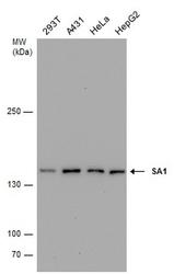
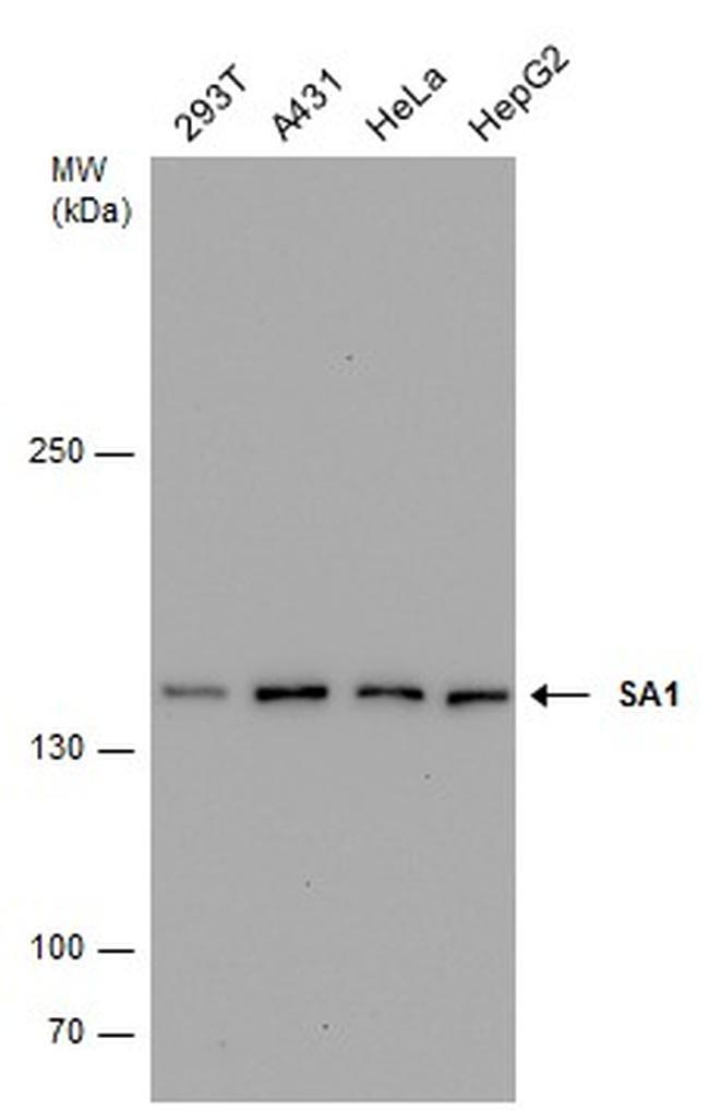
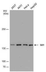
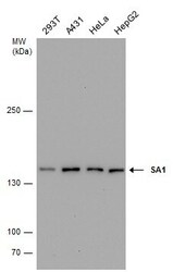
- Western blot analysis of SA1 in whole cell lysate using 30 µg of protein. Samples were separated with 5% SDS-PAGE and incubated with SA1 monoclonal antibody (Product # MA5-27807) using a dilution of 1:1000.
- Submitted by
- Invitrogen Antibodies (provider)
- Main image
- Experimental details
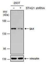
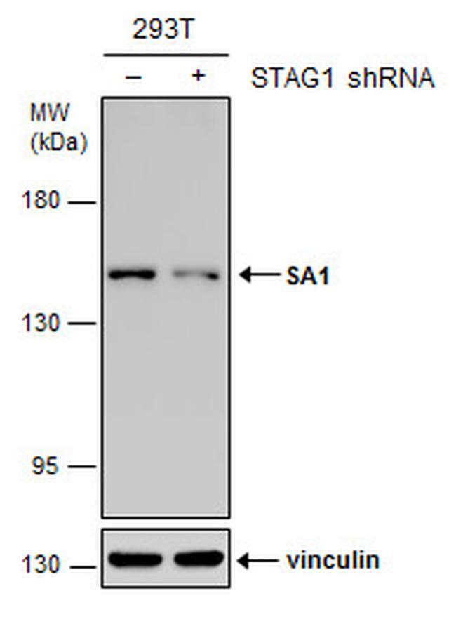
- Western blot analysis of SA1 in non-transfected (-) and transfected (+) 293T cells using 30 µg of protein. Samples were separated with 5% SDS-PAGE and incubated with SA1 monoclonal antibody (Product # MA5-27807) using a dilution of 1:500.
- Submitted by
- Invitrogen Antibodies (provider)
- Main image
- Experimental details
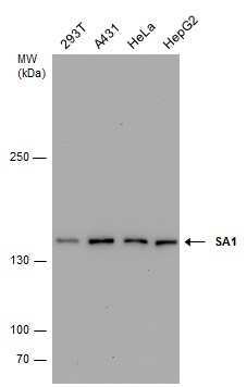
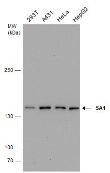
- Western Blot using STAG1 Monoclonal Antibody (GT8810) (Product # MA5-27807). Various whole cell extracts (30 µg) were separated by 5% SDS-PAGE, and the membrane was blotted with STAG1 Polyclonal Antibody STAG1 Monoclonal Antibody (GT8810) (Product # MA5-27807) diluted at 1:1,000.
- Submitted by
- Invitrogen Antibodies (provider)
- Main image
- Experimental details
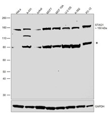
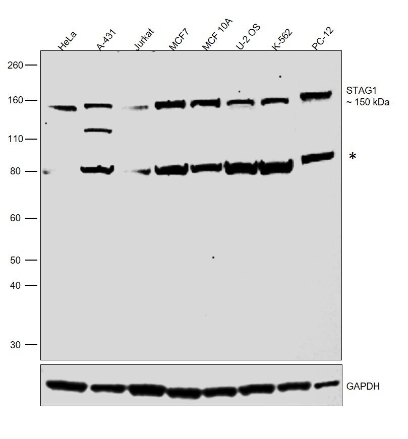
- Western blot was performed using Anti-STAG1 Monoclonal Antibody (GT8810) (Product # MA5-27807) and a 150 kDa band corresponding to Cohesin subunit SA-1 was observed along with an uncharacterized band (*) around 80 kDa. Nuclear enriched extracts (30 µg lysate) of HeLa (Lane 1), A-431 (Lane 2), Jurkat (Lane 3), MCF7 (Lane 4), MCF 10A (Lane 5), U-2 OS (Lane 6), K-562 (Lane 7) and PC-12 (Lane 8) were electrophoresed using NuPAGE™ 4-12% Bis-Tris Protein Gel (Product # NP0322BOX). Resolved proteins were then transferred onto a nitrocellulose membrane (Product # IB23001) by iBlot® 2 Dry Blotting System (Product # IB21001). The blot was probed with the primary antibody (1:1000) and detected by chemiluminescence with Goat anti-Mouse IgG (H+L) Superclonal™ Recombinant Secondary Antibody, HRP (Product # A28177,1:20,000) using the iBright FL 1000 (Product # A32752). Chemiluminescent detection was performed using SuperSignal™ West Pico PLUS Chemiluminescent Substrate (Product # 34580).
- Submitted by
- Invitrogen Antibodies (provider)
- Main image
- Experimental details
- Western Blot using STAG1 Monoclonal Antibody (GT8810) (Product # MA5-27807). Various whole cell extracts (30 µg) were separated by 5% SDS-PAGE, and the membrane was blotted with STAG1 Polyclonal Antibody STAG1 Monoclonal Antibody (GT8810) (Product # MA5-27807) diluted at 1:1,000.
- Submitted by
- Invitrogen Antibodies (provider)
- Main image
- Experimental details
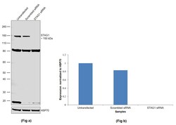
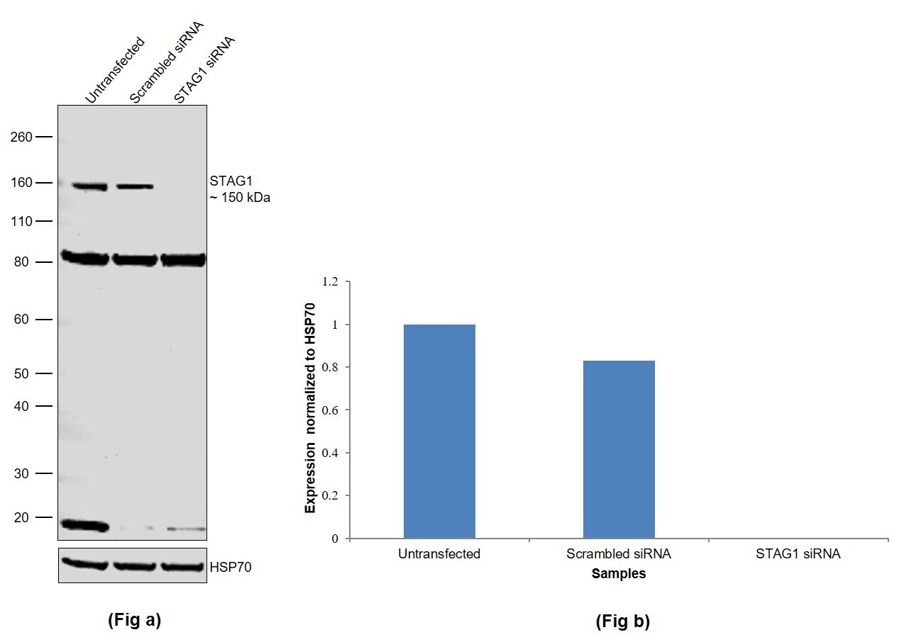
- Knockdown of Cohesin subunit SA-1 was achieved by transfecting MCF 10A with Cohesin subunit SA-1 specific siRNAs (Silencer® select Product # s20075, s20076). Western blot analysis (Fig. a) was performed using Whole cell extracts from the Cohesin subunit SA-1 knockdown cells (lane 3), non-targeting scrambled siRNA transfected cells (lane 2) and untransfected cells (lane 1). The blot was probed with STAG1 Monoclonal Antibody (GT8810) (Product # MA5-27807, 1:1000 ) and Goat anti-Mouse IgG (H+L) Superclonal™ Recombinant Secondary Antibody, HRP (Product # A28177, 1:20,000). Densitometric analysis of this western blot is shown in histogram (Fig. b). Decrease in signal upon siRNA mediated knock down confirms that antibody is specific to Cohesin subunit SA-1.
Supportive validation
- Submitted by
- Invitrogen Antibodies (provider)
- Main image
- Experimental details
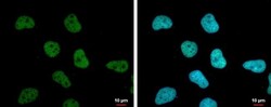
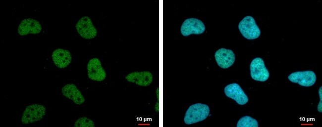
- STAG1 Monoclonal Antibody (GT8810) detects SA1 protein at nucleus by immunofluorescent analysis. Sample: HeLa cells were fixed in 4% paraformaldehyde at RT for 15 min. Green: SA1 protein stained by STAG1 Monoclonal Antibody (GT8810) (Product # MA5-27807) diluted at 1:1,000. Blue: Hoechst 33342 staining. Scale bar = 10 µm.
Supportive validation
- Submitted by
- Invitrogen Antibodies (provider)
- Main image
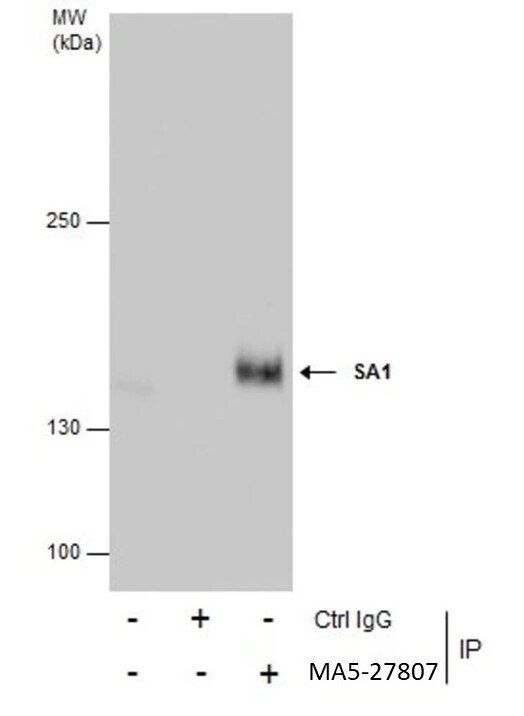
- Experimental details
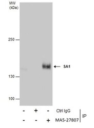
- Immunoprecipitation of SA1 was performed in HeLa whole cell extracts using 5 µg of STAG1 Monoclonal Antibody (GT8810) (Product # MA5-27807). Samples were transferred to a membrane and probed with STAG1 Monoclonal Antibody (GT8810) as a primary antibody and an HRP-conjugated anti-Mouse IgG was used as a secondary antibody.