Explore
 Validate
 Learn
Learn Western blot
 ELISA
ELISAAntibody data
- Antibody Data
- Antigen structure
- References [0]
- Comments [0]
- Validations
- Western blot [1]
- Immunohistochemistry [1]
Submit
Validation data
Reference
Comment
Report error
- Product number
- GTX85615 - Provider product page

- Provider
- GeneTex
- Proper citation
- GeneTex Cat#GTX85615, RRID:AB_10723494
- Product name
- APC1 (phospho Ser377) antibody
- Antibody type
- Polyclonal
- Reactivity
- Human, Mouse
- Host
- Rabbit
No comments: Submit comment
Supportive validation
- Submitted by
- GeneTex (provider)
- Main image
- Experimental details
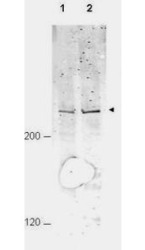
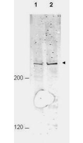
- Western blot of affinity purified anti-APC1 pS377 antibody shows detection of a band ~215 kD corresponding to phosphorylated human APC1 (arrowhead). Lane 1 shows lysate from asynchronous cells. Lane 2 shows lysate from cells treated with nocodazole. While some phosphorylated APC1 is present in untreated cell, the amount of phosphorylated protein is increased in cell preparations arrested in mitosis. Each lane contains approximately 30 ug of HeLa whole cell lysates, separated by 4-8% SDS-PAGE followed by transfer to nitrocellulose. After blocking the membrane was probed with the primary antibody diluted to 1:1000 overnight at 4C followed by washes and reaction with a 1:10000 dilution of IRDye800 conjugated Gt-a-Rabbit IgG [H&L] MX ( for 45 min at room temperature.
Supportive validation
- Submitted by
- GeneTex (provider)
- Main image

- Experimental details
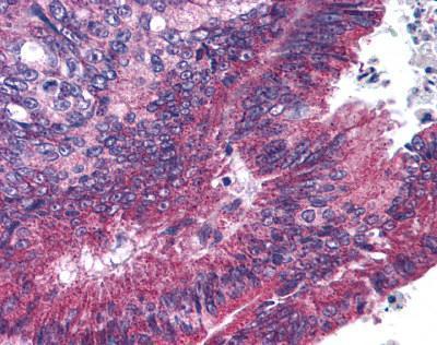
- Human Pancreatic Carcinoma (formalin-fixed, paraffin-embedded) stained with ANAPC1 antibody (GTX85615) at 10 ug/ml followed by biotinylated goat anti-rabbit IgG secondary antibody LS-D1, alkaline phosphatase-streptavidin and chromogen.