 Explore
 Validate
 Learn
Learn Western blot
Antibody data
- Antibody Data
- Antigen structure
- References [2]
- Comments [0]
- Validations
- Western blot [1]
- Immunohistochemistry [1]
Submit
Validation data
Reference
Comment
Report error
- Product number
- PAB9936 - Provider product page

- Provider
- Abnova Corporation
- Proper citation
- Abnova Corporation Cat#PAB9936, RRID:AB_1676184
- Product name
- ANAPC1 (phospho S377) polyclonal antibody
- Antibody type
- Polyclonal
- Description
- Rabbit polyclonal antibody raised against synthetic phosphopeptide of ANAPC1.
- Storage
- Store at 4°C. For long term storage store at -20°C.Aliquot to avoid repeated freezing and thawing.
Submitted references Mitotic regulation of the human anaphase-promoting complex by phosphorylation.
Characterisation of the human APC1, the largest subunit of the anaphase-promoting complex.
Kraft C, Herzog F, Gieffers C, Mechtler K, Hagting A, Pines J, Peters JM
The EMBO journal 2003 Dec 15;22(24):6598-609
The EMBO journal 2003 Dec 15;22(24):6598-609
Characterisation of the human APC1, the largest subunit of the anaphase-promoting complex.
Jörgensen PM, Gräslund S, Betz R, Ståhl S, Larsson C, Höög C
Gene 2001 Jan 10;262(1-2):51-9
Gene 2001 Jan 10;262(1-2):51-9
No comments: Submit comment
Supportive validation
- Submitted by
- Abnova Corporation (provider)
- Main image
- Experimental details
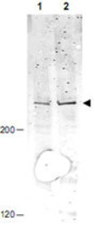
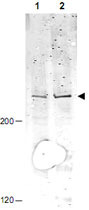
- Western blot using ANAPC1 (phospho S377) polyclonal antibody (Cat # PAB9936) shows detection of a band ~215 KDa corresponding to phosphorylated human ANAPC1 (arrowhead).Lane 1 shows lysate from asynchronous cells.Lane 2 shows lysate from cells treated with nocodazole.While some phosphorylated ANAPC1 is present in untreated cell, the amount of phosphorylated protein is increased in cell preparations arrested in mitosis.Each lane contains approximately 30 ug of HeLa whole cell lysates, separated by 4-8% SDS-PAGE followed by transfer to nitrocellulose.After blocking the membrane was probed with the primary antibody diluted to 1 : 1,000 overnight at 4°C followed by washes and reaction with a 1 : 10,000 dilution of IRDye™800 conjugated Gt-a-Rabbit IgG [H&L] MX for 45 min at room temperature.IRDye™800 fluorescence image was captured using the Odyssey® Infrared Imaging System developed by LI-COR.IRDye is a trademark of LI-COR, Inc.
Supportive validation
- Submitted by
- Abnova Corporation (provider)
- Main image
- Experimental details
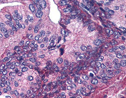
- Immunohistochemistry of ANAPC1 (phospho S377) polyclonal antibody (Cat # PAB9936) was used at 5.0 ug/mL to detect signal in a variety of tissues including multi-human, multi-brain and multi-cancer slides.This image shows moderate positive cytoplasmic and occasional nuclear staining of pancreatic carcinoma cells at 60X.Tissue was formalin-fixed and paraffin embedded.The image shows localization of the antibody as the precipitated red signal, with a hematoxylin purple nuclear counterstain.Personal Communication, Tina Roush, Life Span Biosciences, Seattle, WA.
- Validation comment
- Immunohistochemistry (Formalin/PFA-fixed paraffin-embedded sections)
