Explore
 Validate
 Learn
Learn Western blot
Antibody data
- Antibody Data
- Antigen structure
- References [2]
- Comments [0]
- Validations
- Western blot [1]
- Immunohistochemistry [7]
Submit
Validation data
Reference
Comment
Report error
- Product number
- HPA001352 - Provider product page

- Provider
- Atlas Antibodies
- Proper citation
- Atlas Antibodies Cat#HPA001352, RRID:AB_1078179
- Product name
- Anti-APOA4
- Antibody type
- Polyclonal
- Reactivity
- Human
- Host
- Rabbit
- Conjugate
- Unconjugated
- Antigen sequence
LEGLTFQMKKNAEELKARISASAEELRQRLAPLAE
DVRGNLRGNTEGLQKSLAELGGHLDQQVEEFRRRV
EPYGENFNKALVQQMEQLRQKLGPHAGDVEGHLSF
LEKDLRDKVNSFFSTFKEKESQDKTLSLP- Isotype
- IgG
- Vial size
- 100 µl
- Storage
- Store at +4°C for short term storage. Long time storage is recommended at -20°C.
Submitted references Variance decomposition of protein profiles from antibody arrays using a longitudinal twin model.
Global proteomic profiling reveals altered proteomic signature in schizophrenia serum
Kato BS, Nicholson G, Neiman M, Rantalainen M, Holmes CC, Barrett A, Uhlén M, Nilsson P, Spector TD, Schwenk JM
Proteome science 2011 Nov 17;9:73
Proteome science 2011 Nov 17;9:73
Global proteomic profiling reveals altered proteomic signature in schizophrenia serum
Levin Y, Wang L, Schwarz E, Koethe D, Leweke F, Bahn S
Molecular Psychiatry 2009 June;15(11):1088-1100
Molecular Psychiatry 2009 June;15(11):1088-1100
No comments: Submit comment
Enhanced validation
- Submitted by
- Atlas Antibodies (provider)
- Enhanced method
- Recombinant expression validation
- Main image
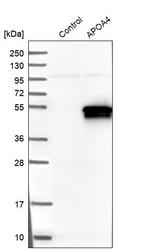
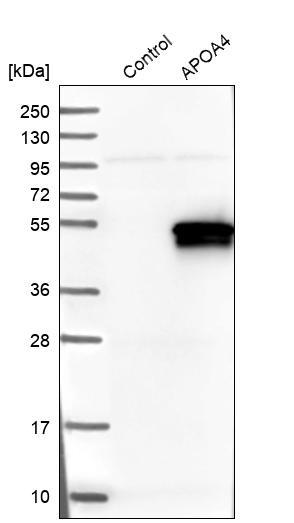
- Experimental details
- Western blot analysis in control (vector only transfected HEK293T lysate) and APOA4 over-expression lysate (Co-expressed with a C-terminal myc-DDK tag (~3.1 kDa) in mammalian HEK293T cells, LY424692).
Enhanced validation
Supportive validation
- Submitted by
- Atlas Antibodies (provider)
- Enhanced method
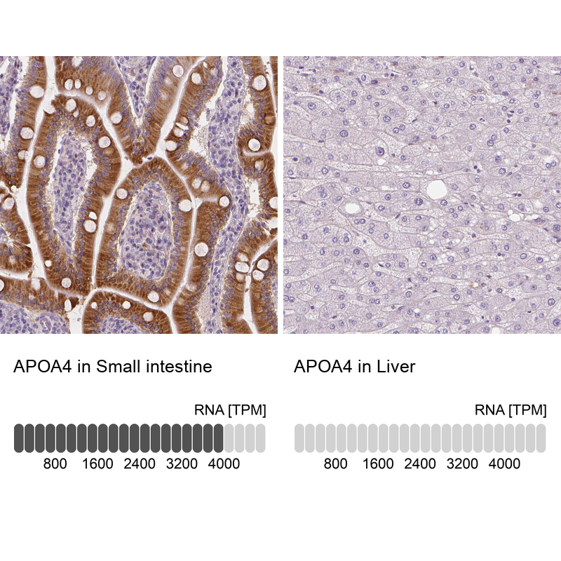
- Orthogonal validation
- Main image
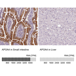
- Experimental details
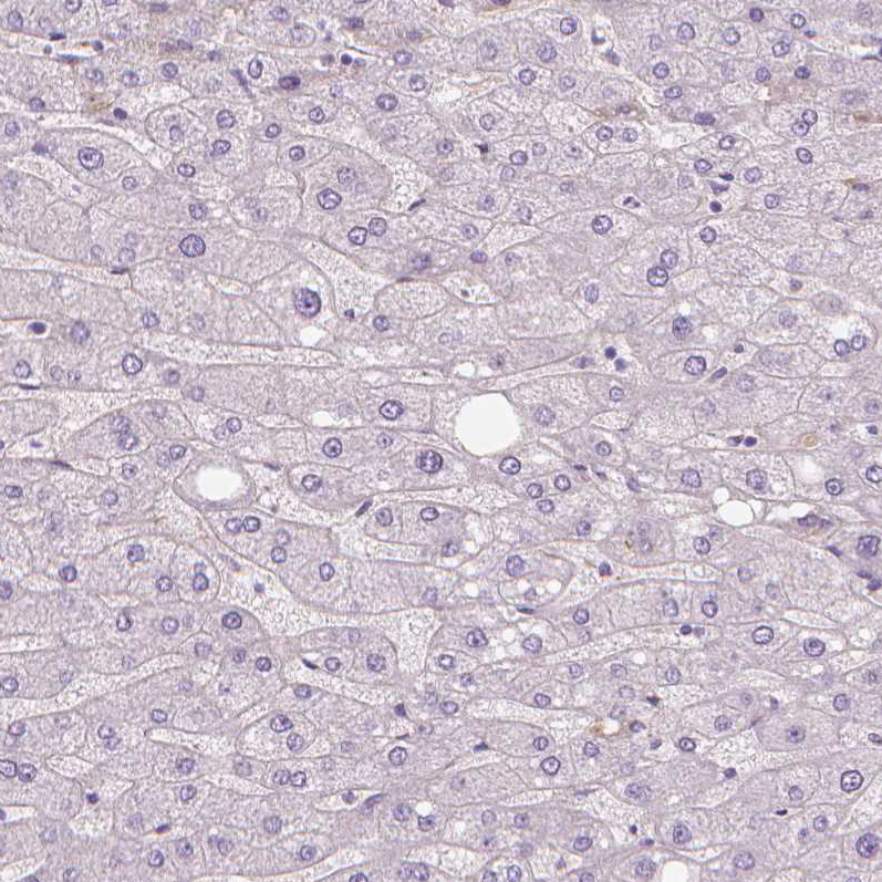
- Immunohistochemistry analysis in human small intestine and liver tissues using HPA001352 antibody. Corresponding APOA4 RNA-seq data are presented for the same tissues.
- Sample type
- HUMAN
Supportive validation
- Submitted by
- Atlas Antibodies (provider)
- Main image
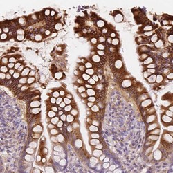
- Experimental details
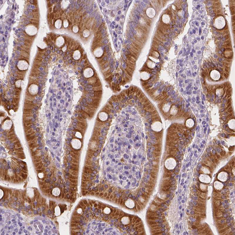
- Immunohistochemical staining of human small intestine shows high expression.
- Sample type
- HUMAN
- Submitted by
- Atlas Antibodies (provider)
- Main image
- Experimental details
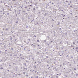
- Immunohistochemical staining of human liver shows low expression as expected.
- Sample type
- HUMAN
- Submitted by
- Atlas Antibodies (provider)
- Main image
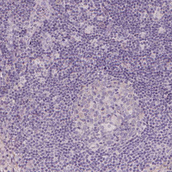
- Experimental details
- Immunohistochemical staining of human lymph node shows no positivity in non-germinal center cells as expected.
- Sample type
- HUMAN

- Submitted by
- Atlas Antibodies (provider)
- Main image
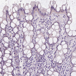
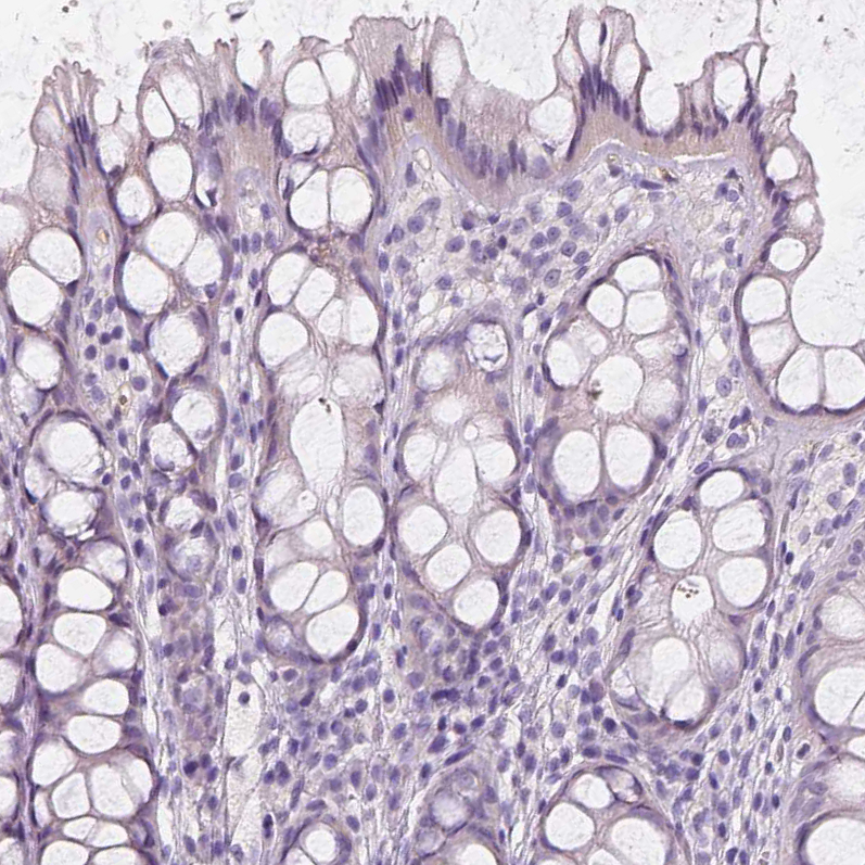
- Experimental details
- Immunohistochemical staining of human colon shows no positivity in glandular cells as expected.
- Sample type
- HUMAN
- Submitted by
- Atlas Antibodies (provider)
- Main image
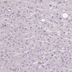
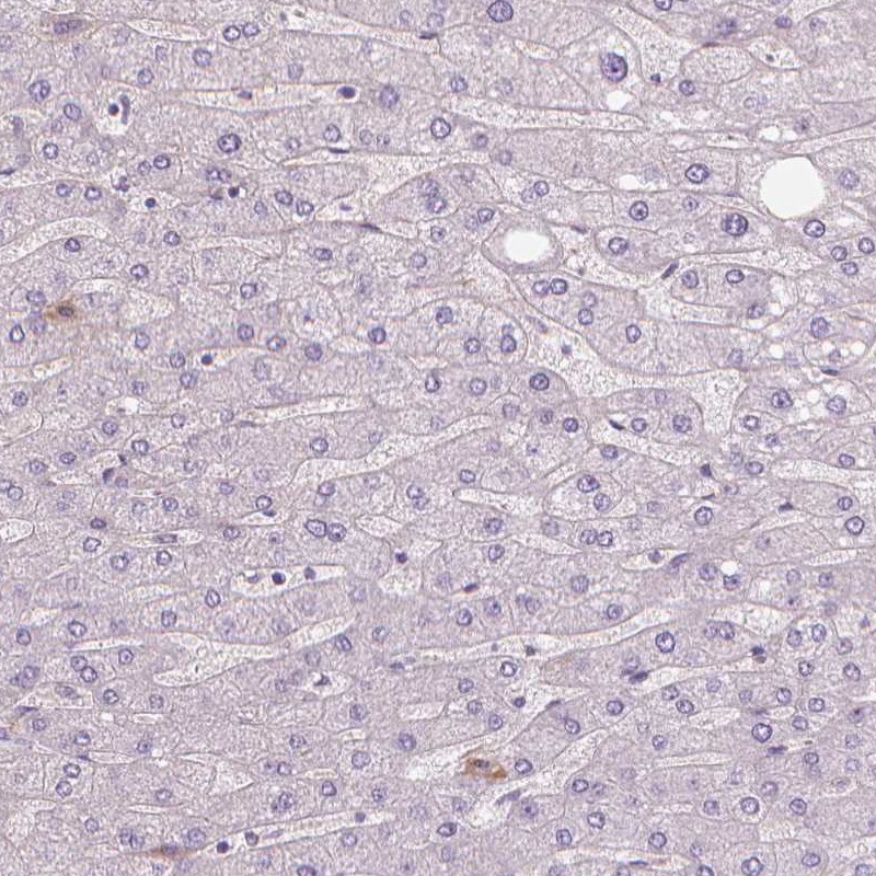
- Experimental details
- Immunohistochemical staining of human liver shows no positivity in hepatocytes as expected.
- Sample type
- HUMAN
- Submitted by
- Atlas Antibodies (provider)
- Main image

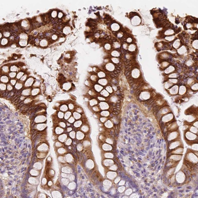
- Experimental details
- Immunohistochemical staining of human small intestine shows strong cytoplasmic positivity in glandular cells.
- Sample type
- HUMAN