 Explore
 Validate
 Learn
LearnMA1-25038
antibody from Invitrogen Antibodies
Targeting: CGA
FSHA, GPA1, GPHa, GPHA1, HCG, LHA, TSHA
 Western blot
 Immunocytochemistry
Antibody data
- Antibody Data
- Antigen structure
- References [1]
- Comments [0]
- Validations
- Immunocytochemistry [1]
Submit
Validation data
Reference
Comment
Report error
- Product number
- MA1-25038 - Provider product page

- Provider
- Invitrogen Antibodies
- Product name
- CGA Monoclonal Antibody (INN-hFSH-132)
- Antibody type
- Monoclonal
- Antigen
- Other
- Description
- MA1-25038 will not react with glycoprotein hormones of various animal species.
- Reactivity
- Human
- Host
- Mouse
- Isotype
- IgG
- Antibody clone number
- INN-hFSH-132
- Vial size
- 250 µg
- Concentration
- 1 mg/mL
- Storage
- Store at 4°C short term. For long term storage, store at -20°C, avoiding freeze/thaw cycles.
Submitted references A high-throughput analysis of the IDH1(R132H) protein expression in pituitary adenomas.
Casar-Borota O, Øystese KA, Sundström M, Melchior L, Popovic V
Pituitary 2016 Aug;19(4):407-14
Pituitary 2016 Aug;19(4):407-14
No comments: Submit comment
Supportive validation
- Submitted by
- Invitrogen Antibodies (provider)
- Main image
- Experimental details
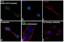
- Immunofluorescence analysis of CGA was performed using 70% confluent log phase BeWo cells treated with Protein Transport Inhibitor (1X, 4hrs). The cells were fixed with 4% paraformaldehyde for 10 minutes, permeabilized with 0.1% Triton™ X-100 for 15 minutes, and blocked with 2% BSA for 45 minutes at room temperature. The cells were labeled with CGA Monoclonal Antibody (INN-hFSH-132) (Product # MA1-25038) at 1:200 dilution in 0.1% BSA, incubated at 4 degree celsius overnight and then labeled with Goat anti-Rabbit IgG (H+L) Highly Cross-Adsorbed Secondary Antibody, Alexa Fluor Plus 488 (Product # A32731), (1:3000 dilution), for 45 minutes at room temperature (Panel a: Green). Nuclei (Panel b:Blue) were stained with ProLong™ Diamond Antifade Mountant with DAPI (Product # P36962). F-actin (Panel c: Red) was stained with Rhodamine Phalloidin (Product # R415, 1:300). Panel d represents the merged image showing cytoplasmic localization. Panel e represents merged image for untreated BeWo cells showing no staining for CGA. Panel f represents control cells with no primary antibody to assess background. The images were captured at 60X magnification.
