Explore
 Validate
 Learn
Learn Western blot
 Immunohistochemistry
Antibody data
- Antibody Data
- Antigen structure
- References [0]
- Comments [0]
- Validations
- Western blot [3]
- Immunocytochemistry [1]
- Immunohistochemistry [9]
Submit
Validation data
Reference
Comment
Report error
- Product number
- HPA020053 - Provider product page

- Provider
- Atlas Antibodies
- Proper citation
- Atlas Antibodies Cat#HPA020053, RRID:AB_1858280
- Product name
- Anti-TRIM29
- Antibody type
- Polyclonal
- Reactivity
- Human, Rat
- Host
- Rabbit
- Conjugate
- Unconjugated
- Antigen sequence
GDDKNSNYFSMDSMEGKRSPYAGLQLGAAKKPPVT
FAEKGELRKSIFSESRKPTVSIMEPGETRRNSYPR
ADTGLFSRSKSGSEEVLCDSCIGNKQKAVKSCLVC
QASFCELHL- Isotype
- IgG
- Vial size
- 100 µl
- Storage
- Store at +4°C for short term storage. Long time storage is recommended at -20°C.
No comments: Submit comment
Supportive validation
- Submitted by
- Atlas Antibodies (provider)
- Main image

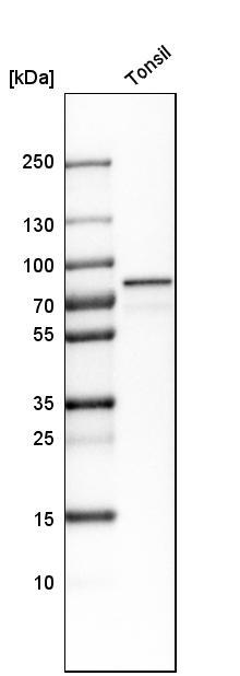
- Experimental details
- Western blot analysis in human tonsil tissue.
- Submitted by
- Atlas Antibodies (provider)
- Main image
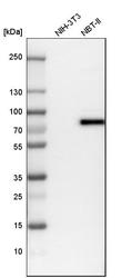
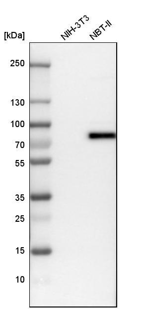
- Experimental details
- Western blot analysis in mouse cell line NIH-3T3 and rat cell line NBT-II.
- Submitted by
- Atlas Antibodies (provider)
- Main image
- Experimental details
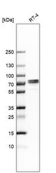
- Western blot analysis in human cell line RT-4.

Supportive validation
- Submitted by
- Atlas Antibodies (provider)
- Main image
- Experimental details
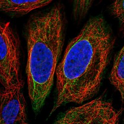
- Immunofluorescent staining of human cell line SiHa shows localization to intermediate filaments.
- Sample type
- HUMAN

Enhanced validation
Supportive validation
- Submitted by
- Atlas Antibodies (provider)
- Enhanced method
- Orthogonal validation
- Main image
- Experimental details
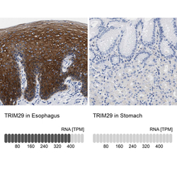
- Immunohistochemistry analysis in human esophagus and stomach tissues using HPA020053 antibody. Corresponding TRIM29 RNA-seq data are presented for the same tissues.
- Sample type
- HUMAN

Supportive validation
- Submitted by
- Atlas Antibodies (provider)
- Main image

- Experimental details
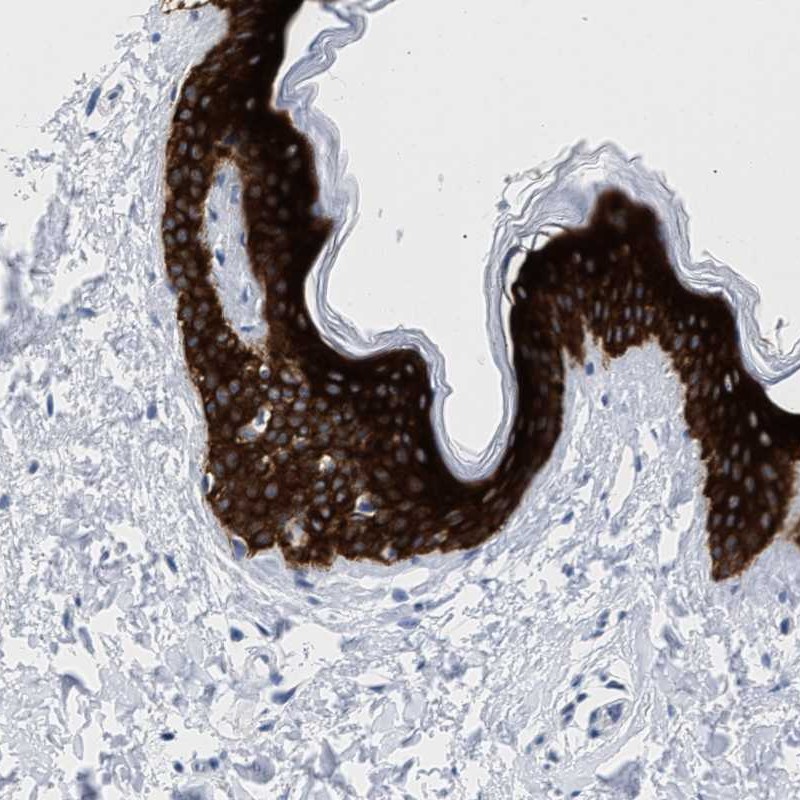
- Immunohistochemical staining of human skin shows strong cytoplasmic positivity in epidermal cells.
- Submitted by
- Atlas Antibodies (provider)
- Main image
- Experimental details
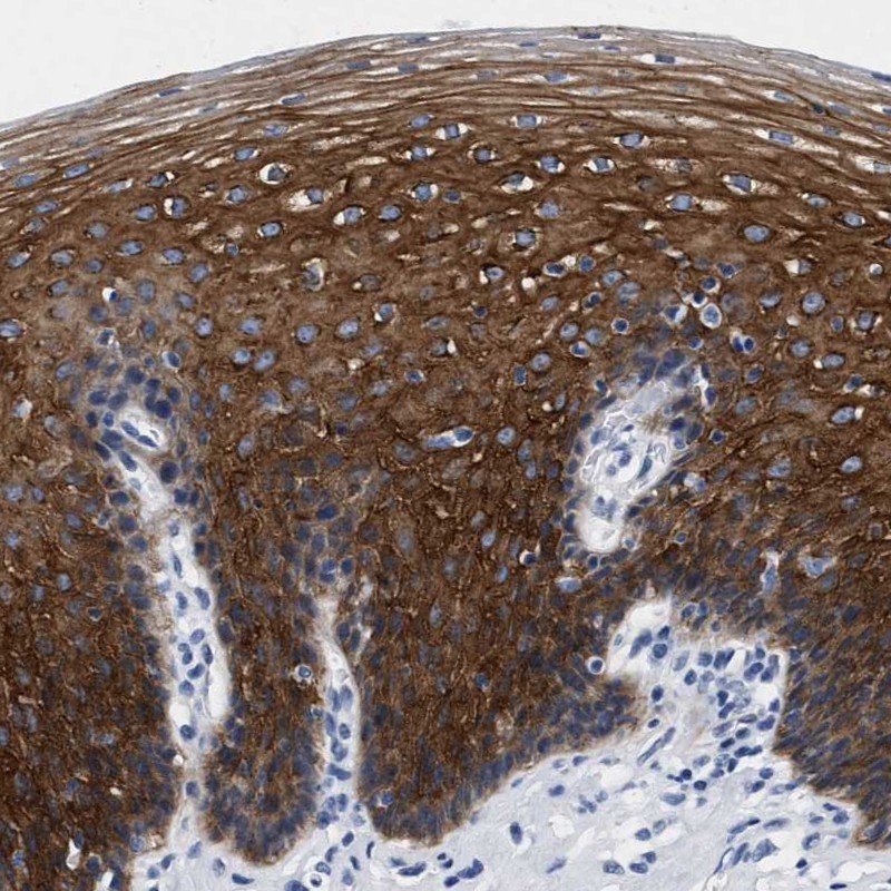
- Immunohistochemical staining of human esophagus shows high expression.
- Sample type
- HUMAN
- Submitted by
- Atlas Antibodies (provider)
- Main image

- Experimental details
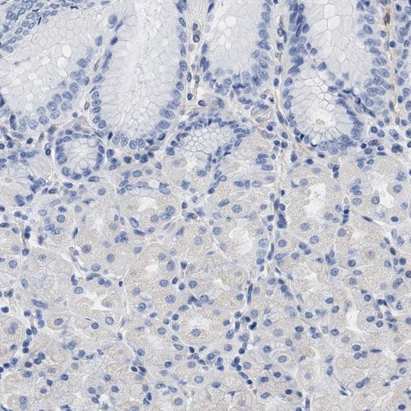
- Immunohistochemical staining of human stomach shows low expression as expected.
- Sample type
- HUMAN
- Submitted by
- Atlas Antibodies (provider)
- Main image

- Experimental details
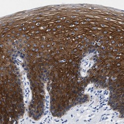
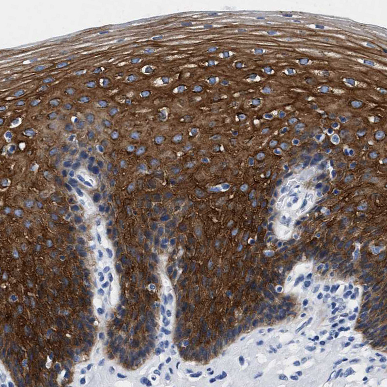
- Immunohistochemical staining of human esophagus shows cytoplasmic positivity in squamous epithelial cells.
- Sample type
- HUMAN
- Submitted by
- Atlas Antibodies (provider)
- Main image

- Experimental details
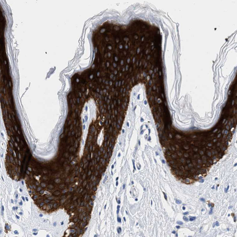
- Immunohistochemical staining of human skin shows cytoplasmic positivity in epidermal cells.
- Sample type
- HUMAN
- Submitted by
- Atlas Antibodies (provider)
- Main image
- Experimental details
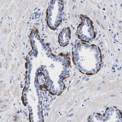
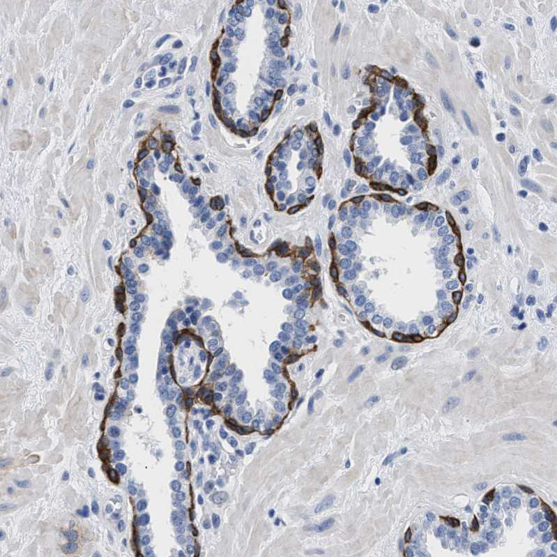
- Immunohistochemical staining of human prostate shows cytoplasmic positivity in basal layer of glandular cells.
- Sample type
- HUMAN
- Submitted by
- Atlas Antibodies (provider)
- Main image

- Experimental details
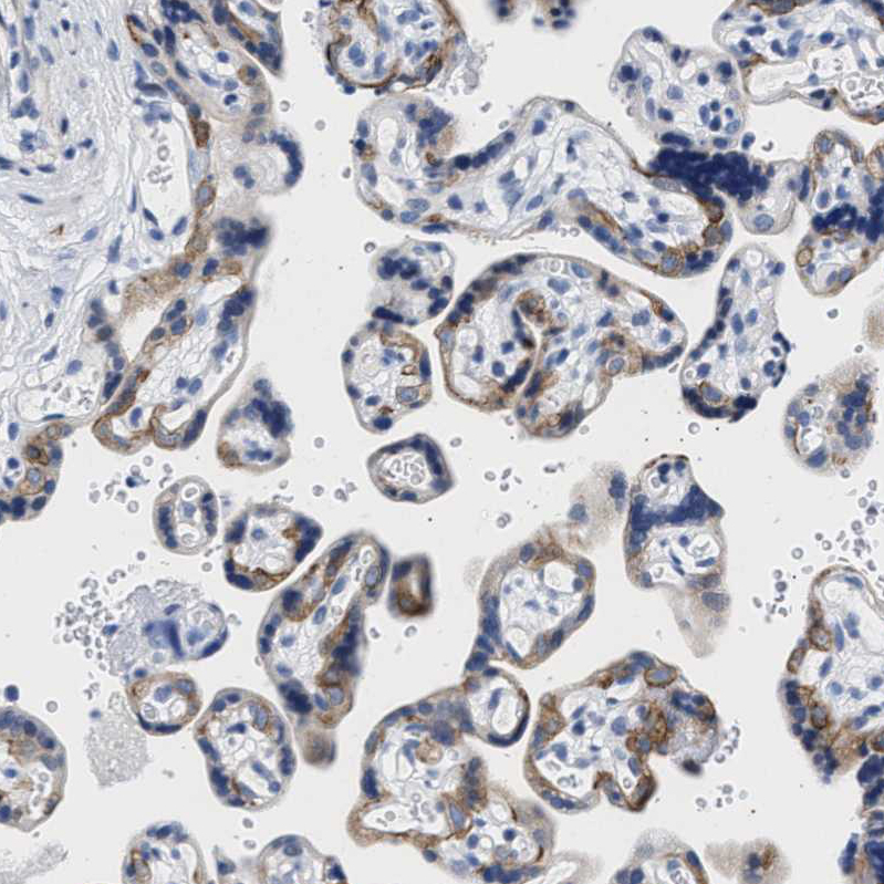
- Immunohistochemical staining of human placenta shows cytoplasmic positivity in trophoblastic cells.
- Sample type
- HUMAN
- Submitted by
- Atlas Antibodies (provider)
- Main image

- Experimental details
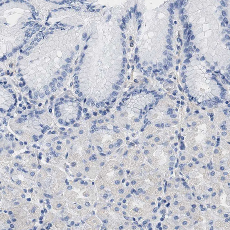
- Immunohistochemical staining of human stomach shows no positivity in glandular cells as expected.
- Sample type
- HUMAN