 Explore
 Validate
 Learn
Learn Western blot
 Immunocytochemistry
Antibody data
- Antibody Data
- Antigen structure
- References [2]
- Comments [0]
- Validations
- Western blot [2]
- Immunoprecipitation [1]
- Immunohistochemistry [1]
Submit
Validation data
Reference
Comment
Report error
- Product number
- NBP1-32695 - Provider product page

- Provider
- Novus Biologicals
- Proper citation
- Novus Cat#NBP1-32695, RRID:AB_2241808
- Product name
- Rabbit Polyclonal 14-3-3 epsilon Antibody
- Antibody type
- Polyclonal
- Description
- Immunogen affinity purified.
- Reactivity
- Human, Mouse
- Host
- Rabbit
- Isotype
- IgG
- Vial size
- 0.1 ml
- Storage
- Aliquot and store at -20C or -80C. Avoid freeze-thaw cycles.
Submitted references A novel inhibitory anti-invasive MAb isolated using phenotypic screening highlights AnxA6 as a functionally relevant target protein in pancreatic cancer.
Quantitative label-free mass spectrometry analysis of formalin-fixed, paraffin-embedded tissue representing the invasive cutaneous malignant melanoma proteome.
O'Sullivan D, Dowling P, Joyce H, McAuley E, McCann A, Henry M, McGovern B, Barham P, Kelleher FC, Murphy J, Kennedy S, Swan N, Moriarty M, Clynes M, Larkin A
British journal of cancer 2017 Oct 24;117(9):1326-1335
British journal of cancer 2017 Oct 24;117(9):1326-1335
Quantitative label-free mass spectrometry analysis of formalin-fixed, paraffin-embedded tissue representing the invasive cutaneous malignant melanoma proteome.
Dowling P, Moran B, McAuley E, Meleady P, Henry M, Clynes M, McMenamin M, Leonard N, Monks M, Wynne B, Ormond P, Larkin A
Oncology letters 2016 Nov;12(5):3296-3304
Oncology letters 2016 Nov;12(5):3296-3304
No comments: Submit comment
Supportive validation
- Submitted by
- Novus Biologicals (provider)
- Main image
- Experimental details
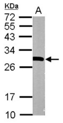
- Western Blot: 14-3-3 epsilon Antibody [NBP1-32695] - Sample (20 ug of whole cell lysate) A: mouse brain 12% SDS PAGE; antibody diluted at 1:10000.

- Submitted by
- Novus Biologicals (provider)
- Main image
- Experimental details
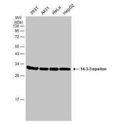
- Western Blot: 14-3-3 epsilon Antibody [NBP1-32695] - Various whole cell extracts (30 ug) were separated by 12% SDS-PAGE, and the membrane was blotted with 14-3-3 epsilon antibody [N1C3] diluted at 1:5000. The HRP-conjugated anti-rabbit IgG antibody (NBP2-19301) was used to detect the primary antibody

Supportive validation
- Submitted by
- Novus Biologicals (provider)
- Main image

- Experimental details
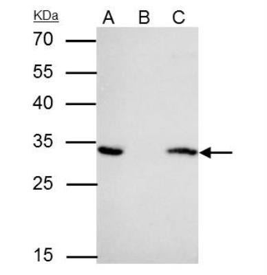
- Immunoprecipitation: 14-3-3 epsilon Antibody [NBP1-32695] - IP samples: HeLa whole cell extract A. 40 ug HeLa whole cell extract B. Control with 4 ug of preimmune Rabbit IgG C. Immunoprecipitation of YWHAE protein by 4 ug 14-3-3 epsilon antibody [N1C3] (NBP1-32695) 12 % SDS-PAGE The immunoprecipitated YWHAE protein was detected by 14-3-3 epsilon antibody [N1C3] (NBP1-32695) diluted at 1:1000. [EasyBlot anti-rabbit IgG was used as a secondary reagent]
Supportive validation
- Submitted by
- Novus Biologicals (provider)
- Main image
- Experimental details
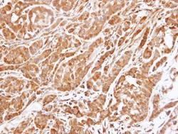
- Immunohistochemistry-Paraffin: 14-3-3 epsilon Antibody [NBP1-32695] - Paraffin-embedded A549 xenograft , using 14-3-3 epsilon antibody at 1:500 dilution.
