Explore
 Validate
 Learn
Learn Western blot
Antibody data
- Antibody Data
- Antigen structure
- References [2]
- Comments [0]
- Validations
- Western blot [1]
- Immunocytochemistry [1]
- Immunohistochemistry [1]
- Flow cytometry [5]
Submit
Validation data
Reference
Comment
Report error
- Product number
- MA1-16874 - Provider product page

- Provider
- Invitrogen Antibodies
- Product name
- Blimp-1 Monoclonal Antibody (3H2-E8)
- Antibody type
- Monoclonal
- Antigen
- Other
- Description
- In Western Blot, this antibody recognizes a band at ~98 kDa and may recognize one at ~80 kDa (the beta form).
- Antibody clone number
- 3H2-E8
- Concentration
- 1 mg/mL
Submitted references bFGF signaling-mediated reprogramming of porcine primordial germ cells.
Critical function of AP-2 gamma/TCFAP2C in mouse embryonic germ cell maintenance.
Zhang Y, Ma J, Li H, Lv J, Wei R, Cong Y, Liu Z
Cell and tissue research 2016 May;364(2):429-41
Cell and tissue research 2016 May;364(2):429-41
Critical function of AP-2 gamma/TCFAP2C in mouse embryonic germ cell maintenance.
Weber S, Eckert D, Nettersheim D, Gillis AJ, Schäfer S, Kuckenberg P, Ehlermann J, Werling U, Biermann K, Looijenga LH, Schorle H
Biology of reproduction 2010 Jan;82(1):214-23
Biology of reproduction 2010 Jan;82(1):214-23
No comments: Submit comment
Supportive validation
- Submitted by
- Invitrogen Antibodies (provider)
- Main image
- Experimental details
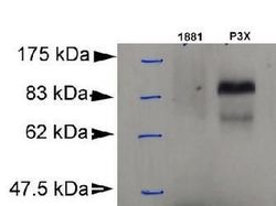
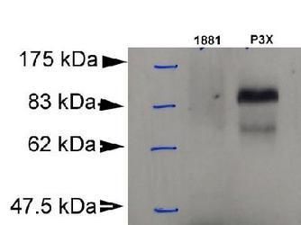
- Western Blot detection of Blimp-1 in murine plasmacytoma cell lysate (P3X) using Product # MA1-16874. 1881: murine pre-B cell lysate (negative control).
Supportive validation
- Submitted by
- Invitrogen Antibodies (provider)
- Main image
- Experimental details
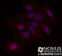
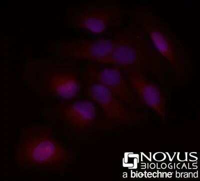
- Immunocytochemistry analysis of Blimp-1 in HeLa cells fixed for 10 minutes using 10% formalin and then permeabilized for 5 minutes using 1X PBS + 0.05% Triton-X100. Samples were incubated in Blimp-1 monoclonal antibody (Product # MA1-16874) using a dilution of 20 µg/mL for 1 hour at room temperature. Antibody conjugated to DyLight 550. Nuclei were counterstained with DAPI (Blue). Cells were imaged using a 40X objective.
Supportive validation
- Submitted by
- Invitrogen Antibodies (provider)
- Main image

- Experimental details
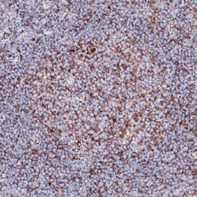
- Immunohistochemical analysis of Blimp-1 in paraffin-embedded human tonsil tissue. Samples were incubated in Blimp-1 monoclonal antibody (Product # MA1-16874) using a dilution of 0.0763888888888889. Detection is completed through DAB and Counterstained by hematoxylin; labeling is predominantly in germinal centers.
Supportive validation
- Submitted by
- Invitrogen Antibodies (provider)
- Main image
- Experimental details
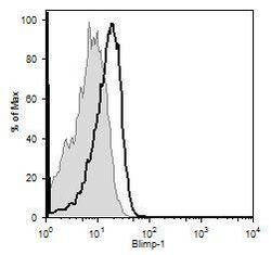
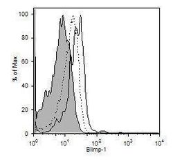
- Blimp-1 expression by CD19+ (thick black line) or CD8+ (thin, shaded line).
- Submitted by
- Invitrogen Antibodies (provider)
- Main image
- Experimental details
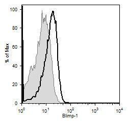
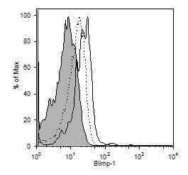
- Blimp-1 expression by IL-10+ (black line), IL-10- (dotted line) or CD8+ (thin, shaded line) cells.
- Submitted by
- Invitrogen Antibodies (provider)
- Main image
- Experimental details
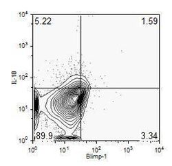
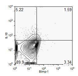
- IL-10+Blimp-1-, IL-10+Blimp-1+, IL-10-Blimp-1+ and IL-10-Blimp-1- cells, % total living single splenic CD19+ cells of 3 total mice.
- Submitted by
- Invitrogen Antibodies (provider)
- Main image
- Experimental details
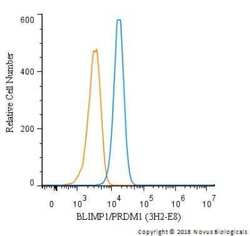
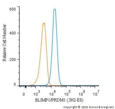
- Flow cytometry of Blimp-1 in U266 cells. Samples were incubated in Blimp-1 monoclonal antibody (Product # MA1-16874) using a dilution of 2.5 µg/mL for 30 minutes at room temperature followed by mouse F(ab)2 IgG (H+L) APC-conjugated secondary antibody. Antibody (blue) and a matched isotype control (orange). Cells were fixed with 4% PFA and permeabilized with 0.1% Saponin.
- Submitted by
- Invitrogen Antibodies (provider)
- Main image
- Experimental details
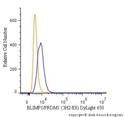
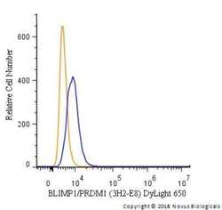
- Flow cytometry of Blimp-1 in A431 cells. Samples were incubated in Blimp-1 monoclonal antibody (Product # MA1-16874) using a dilution of 5 µg/mL for 30 minutes at room temperature. Antibody (blue) and a matched isotype control (orange). Cells were fixed with 4% PFA and then permeabilized with 0.1% saponin. Both antibodies were conjugated to DyLight 650.