Explore
 Validate
 Learn
LearnHPA020559
antibody from Atlas Antibodies
Targeting: SERBP1
CGI-55, CHD3IP, DKFZP564M2423, HABP4L, PAI-RBP1, PAIRBP1
 Western blot
Antibody data
- Antibody Data
- Antigen structure
- References [2]
- Comments [0]
- Validations
- Western blot [2]
- Immunocytochemistry [1]
- Immunohistochemistry [5]
Submit
Validation data
Reference
Comment
Report error
- Product number
- HPA020559 - Provider product page

- Provider
- Atlas Antibodies
- Proper citation
- Atlas Antibodies Cat#HPA020559, RRID:AB_1856701
- Product name
- Anti-SERBP1
- Antibody type
- Polyclonal
- Reactivity
- Human, Mouse, Rat
- Host
- Rabbit
- Conjugate
- Unconjugated
- Antigen sequence
GHLQEGFGCVVTNRFDQLFDDESDPFEVLKAAENK
KKEAGGGGVGGPGAKSAAQAAAQTNSNAAGKQLRK
ESQKDRKNPLPPSVGVVDKKEETQPPVALKKEGIR
RVGRRPDQQLQGEGKIIDRRPERRPPRERRFE- Isotype
- IgG
- Vial size
- 100 µl
- Storage
- Store at +4°C for short term storage. Long time storage is recommended at -20°C.
Submitted references Matrix remodeling stimulates stromal autophagy, “fueling” cancer cell mitochondrial metabolism and metastasis
Immunofluorescence and fluorescent-protein tagging show high correlation for protein localization in mammalian cells
Castello-Cros R, Bonnuccelli G, Molchansky A, Capozza F, Witkiewicz A, Birbe R, Howell A, Pestell R, Whitaker-Menezes D, Sotgia F, Lisanti M
Cell Cycle 2014 November;10(12):2021-2034
Cell Cycle 2014 November;10(12):2021-2034
Immunofluorescence and fluorescent-protein tagging show high correlation for protein localization in mammalian cells
Stadler C, Rexhepaj E, Singan V, Murphy R, Pepperkok R, Uhlén M, Simpson J, Lundberg E
Nature Methods 2013 February;10(4):315-323
Nature Methods 2013 February;10(4):315-323
No comments: Submit comment
Supportive validation
- Submitted by
- Atlas Antibodies (provider)
- Main image

- Experimental details
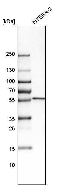
- Western blot analysis in human cell line NTERA-2.
- Submitted by
- Atlas Antibodies (provider)
- Main image
- Experimental details
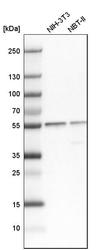
- Western blot analysis in mouse cell line NIH-3T3 and rat cell line NBT-II.
Supportive validation
- Submitted by
- Atlas Antibodies (provider)
- Main image
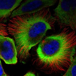
- Experimental details
- Immunofluorescent staining of human cell line U-2 OS shows localization to cytosol.
- Sample type
- HUMAN

Supportive validation
- Submitted by
- Atlas Antibodies (provider)
- Main image
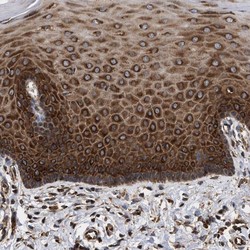
- Experimental details
- Immunohistochemical staining of human cervix, uterine shows strong cytoplasmic positivity in squamous epithelial cells.

- Submitted by
- Atlas Antibodies (provider)
- Main image
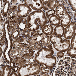
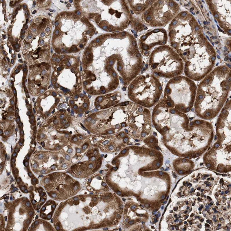
- Experimental details
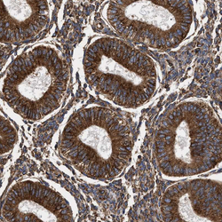
- Immunohistochemical staining of human kidney shows strong cytoplasmic positivity in cells in tubules.
- Sample type
- HUMAN
- Submitted by
- Atlas Antibodies (provider)
- Main image
- Experimental details
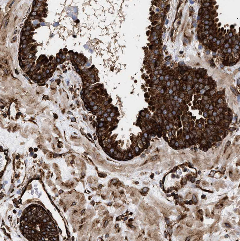
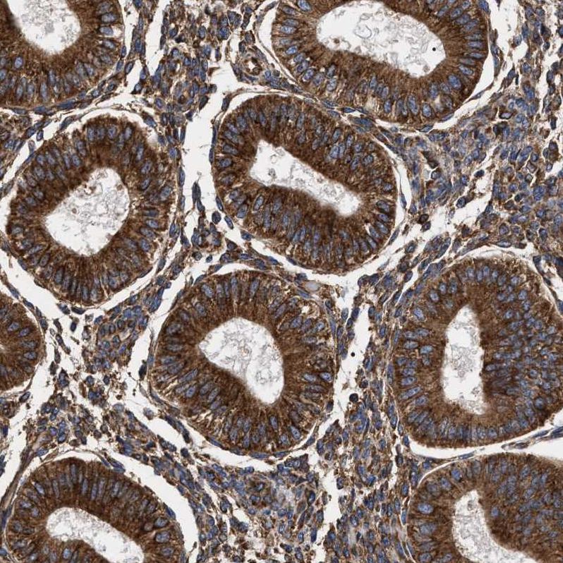
- Immunohistochemical staining of human endometrium shows strong cytoplasmic positivity in glandular cells.
- Sample type
- HUMAN
- Submitted by
- Atlas Antibodies (provider)
- Main image
- Experimental details
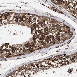
- Immunohistochemical staining of human testis shows strong cytoplasmic positivity in cells in seminiferous ducts.
- Sample type
- HUMAN

- Submitted by
- Atlas Antibodies (provider)
- Main image
- Experimental details
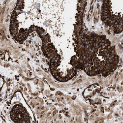
- Immunohistochemical staining of human prostate shows strong cytoplasmic positivity in glandular cells.
- Sample type
- HUMAN