 Explore
 Validate
 Learn
Learn Western blot
Antibody data
- Antibody Data
- Antigen structure
- References [1]
- Comments [0]
- Validations
- Western blot [2]
- Immunocytochemistry [2]
- Immunohistochemistry [4]
- Flow cytometry [1]
- Other assay [1]
Submit
Validation data
Reference
Comment
Report error
- Product number
- MA5-32221 - Provider product page

- Provider
- Invitrogen Antibodies
- Product name
- ATG7 Recombinant Rabbit Monoclonal Antibody (SC06-30)
- Antibody type
- Monoclonal
- Antigen
- Synthetic peptide
- Description
- Recombinant rabbit monoclonal antibodies are produced using in vitro expression systems. The expression systems are developed by cloning in the specific antibody DNA sequences from immunoreactive rabbits. Then, individual clones are screened to select the best candidates for production. The advantages of using recombinant rabbit monoclonal antibodies include: better specificity and sensitivity, lot-to-lot consistency, animal origin-free formulations, and broader immunoreactivity to diverse targets due to larger rabbit immune repertoire.
- Reactivity
- Human
- Host
- Rabbit
- Isotype
- IgG
- Antibody clone number
- SC06-30
- Vial size
- 100 µL
- Concentration
- 1 mg/mL
- Storage
- Store at 4°C short term. For long term storage, store at -20°C, avoiding freeze/thaw cycles.
Submitted references lnc-HOTAIR predicts hepatocellular carcinoma in chronic hepatitis C genotype 4 following direct-acting antivirals therapy.
El-Khazragy N, Elshimy AA, Hassan SS, Shaaban MH, Bayoumi AH, El Magdoub HM, Ghozy S, Gaballah A, Aboelhussein MM, Abou Gabal HH, Bannunah AM, Mansy AE
Molecular carcinogenesis 2020 Dec;59(12):1382-1391
Molecular carcinogenesis 2020 Dec;59(12):1382-1391
No comments: Submit comment
Supportive validation
- Submitted by
- Invitrogen Antibodies (provider)
- Main image

- Experimental details
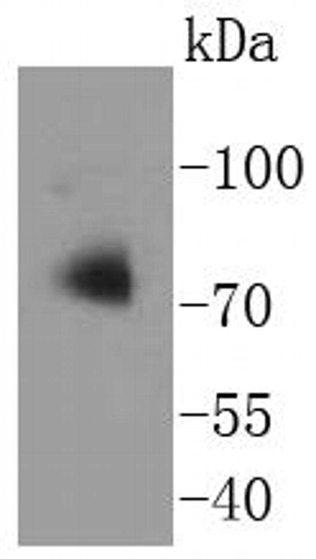
- Western blot analysis of ATG7 in Jurkat cell lysates using a ATG7 Monoclonal antibody (Product # MA5-32221) at a dilution of 1:1,000.
- Submitted by
- Invitrogen Antibodies (provider)
- Main image
- Experimental details
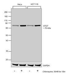
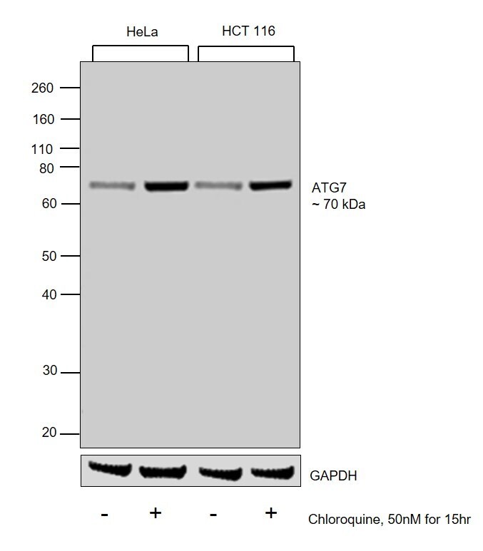
- Western blot was performed using Anti-ATG7 Recombinant Rabbit Monoclonal Antibody (SC06-30) (Product # MA5-32221) and a 70 kDa band corresponding to ATG7 was observed across cell lines tested and increased upon Chloroquine treatment. Whole cell extracts (30 µg lysate) of HeLa (Lane 1), HeLa treated with Chloroquine (50nM for 15 hr) (Lane 2), HCT 116 (Lane 3) and HCT 116 treated with Chloroquine (50nM for 15 hr) (Lane 4) were electrophoresed using Novex® NuPAGE® 12 % Bis-Tris gel (Product # NP0342BOX). Resolved proteins were then transferred onto a nitrocellulose membrane (Product # IB23001) by iBlot® 2 Dry Blotting System (Product # IB21001). The blot was probed with the primary antibody (1:1000 dilution) and detected by chemiluminescence with Goat anti-Rabbit IgG (H+L), Superclonal™ Recombinant Secondary Antibody, HRP (Product # A27036, 1:4000 dilution) using the iBright FL 1000 (Product # A32752). Chemiluminescent detection was performed using Novex® ECL Chemiluminescent Substrate Reagent Kit (Product # WP20005).
Supportive validation
- Submitted by
- Invitrogen Antibodies (provider)
- Main image
- Experimental details
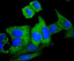
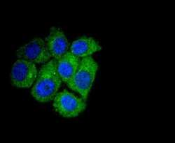
- Immunocytochemical analysis of ATG7 in Hela cells using a ATG7 Monoclonal antibody (Product # MA5-32221) as seen in green. The nuclear counter stain is DAPI (blue). Cells were fixed in paraformaldehyde, permeabilised with 0.25% Triton X100/PBS.

- Submitted by
- Invitrogen Antibodies (provider)
- Main image
- Experimental details
- Immunocytochemical analysis of ATG7 in HepG2 cells using a ATG7 Monoclonal antibody (Product # MA5-32221) as seen in green. The nuclear counter stain is DAPI (blue). Cells were fixed in paraformaldehyde, permeabilised with 0.25% Triton X100/PBS.

Supportive validation
- Submitted by
- Invitrogen Antibodies (provider)
- Main image
- Experimental details
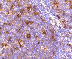
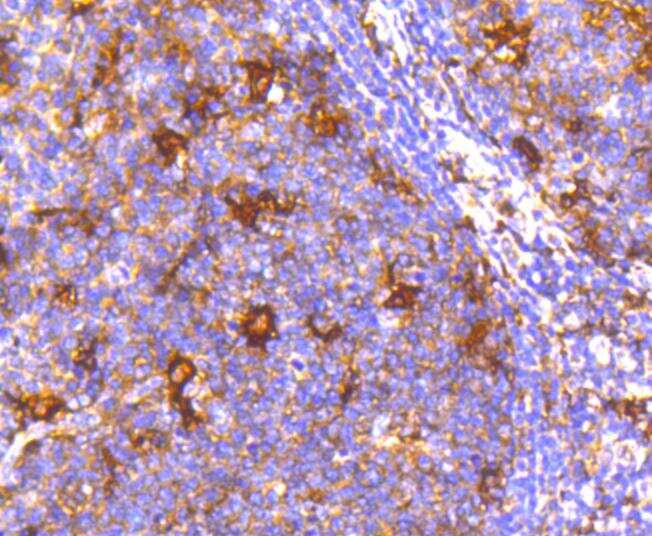
- Immunohistochemical analysis of ATG7 of paraffin-embedded Human tonsil tissue using a ATG7 Monoclonal antibody (Product #MA5-32221). Counter stained with hematoxylin.
- Submitted by
- Invitrogen Antibodies (provider)
- Main image
- Experimental details
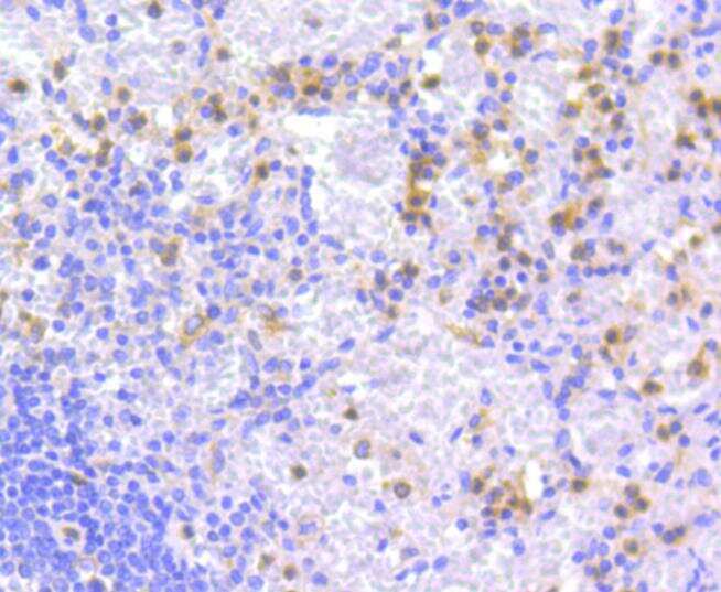
- Immunohistochemical analysis of ATG7 of paraffin-embedded Human spleen tissue using a ATG7 Monoclonal antibody (Product #MA5-32221). Counter stained with hematoxylin.
- Submitted by
- Invitrogen Antibodies (provider)
- Main image
- Experimental details
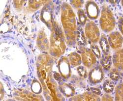
- Immunohistochemical analysis of ATG7 of paraffin-embedded Human kidney tissue using a ATG7 Monoclonal antibody (Product #MA5-32221). Counter stained with hematoxylin.

- Submitted by
- Invitrogen Antibodies (provider)
- Main image

- Experimental details
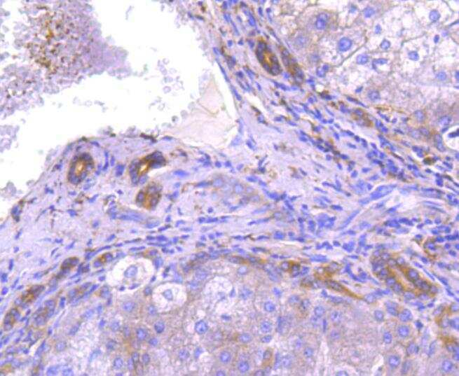
- Immunohistochemical analysis of ATG7 of paraffin-embedded Mouse brain tissue using a ATG7 Monoclonal antibody (Product #MA5-32221). Counter stained with hematoxylin.
Supportive validation
- Submitted by
- Invitrogen Antibodies (provider)
- Main image
- Experimental details
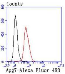
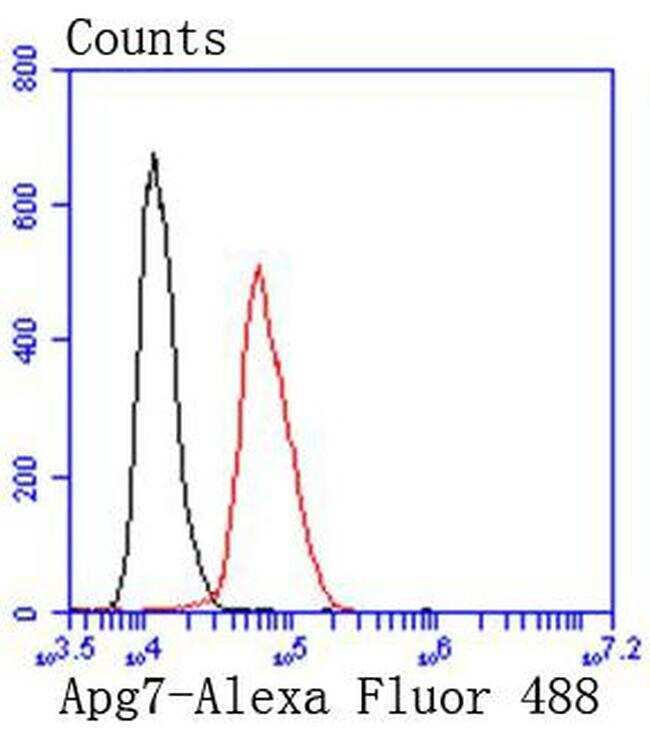
- Flow Cytometric analysis of ATG7 in Hela cells using a ATG7 Monoclonal Antibody (Product # MA5-32221) at a dilution of 1:50, as seen in red compared with an unlabelled control (cells without incubation with primary antibody; black). Alexa Fluor 488-conjugated goat anti rabbit IgG was used as the secondary antibody.
Supportive validation
- Submitted by
- Invitrogen Antibodies (provider)
- Main image
- Experimental details
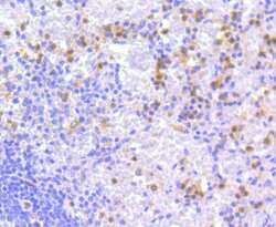
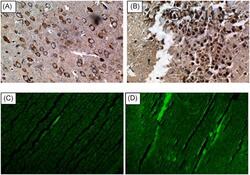
- 4 Figure Immunohistochemically staining of ATG-7 protein in HCC following DAAs therapy in HCV patients the representative image shows the proportion of ATG-7 staining immune-positive cells; (A) 20%-40% HCC-positive cells in patients with lnc-HOTAIR expression levels
