Explore
 Validate
 Learn
Learn Western blot
Antibody data
- Antibody Data
- Antigen structure
- References [2]
- Comments [0]
- Validations
- Western blot [4]
- Immunocytochemistry [1]
- Immunoprecipitation [1]
- Immunohistochemistry [1]
Submit
Validation data
Reference
Comment
Report error
- Product number
- GTX114541 - Provider product page

- Provider
- GeneTex
- Proper citation
- GeneTex Cat#GTX114541, RRID:AB_10619599
- Product name
- MAFG antibody
- Antibody type
- Polyclonal
- Reactivity
- Human, Mouse, Rat
- Host
- Rabbit
Submitted references miR-128 Is Implicated in Stress Responses by Targeting MAFG in Skeletal Muscle Cells.
MAFG is a transcriptional repressor of bile acid synthesis and metabolism.
Caggiano R, Cattaneo F, Moltedo O, Esposito G, Perrino C, Trimarco B, Ammendola R, Faraonio R
Oxidative medicine and cellular longevity 2017;2017:9308310
Oxidative medicine and cellular longevity 2017;2017:9308310
MAFG is a transcriptional repressor of bile acid synthesis and metabolism.
de Aguiar Vallim TQ, Tarling EJ, Ahn H, Hagey LR, Romanoski CE, Lee RG, Graham MJ, Motohashi H, Yamamoto M, Edwards PA
Cell metabolism 2015 Feb 3;21(2):298-311
Cell metabolism 2015 Feb 3;21(2):298-311
No comments: Submit comment
Supportive validation
- Submitted by
- GeneTex (provider)
- Main image
- Experimental details
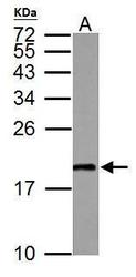
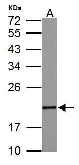
- MAFG antibody detects MAFG protein by western blot analysis.A.50 ?g mouse muscle lysate/extract 12% SDS-PAGEMAFG antibody (GTX114541) dilution: 1:1000 The HRP-conjugated anti-rabbit IgG antibody (GTX213110-01) was used to detect the primary antibody.
- Submitted by
- GeneTex (provider)
- Main image
- Experimental details
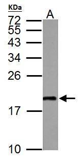
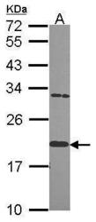
- MAFG antibody detects MAFG protein by western blot analysis.A.50 ?g rat muscle lysate/extract 12% SDS-PAGEMAFG antibody (GTX114541) dilution: 1:1000 The HRP-conjugated anti-rabbit IgG antibody (GTX213110-01) was used to detect the primary antibody.
- Submitted by
- GeneTex (provider)
- Main image
- Experimental details
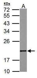
- Sample (30 ug of whole cell lysate) A: Hela 12% SDS PAGE GTX114541 diluted at 1:1000
- Validation comment
- WB

- Submitted by
- GeneTex (provider)
- Main image
- Experimental details
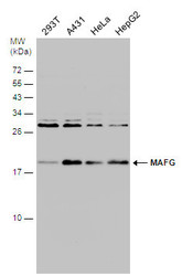
- Various whole cell extracts (30 ?g) were separated by 15% SDS-PAGE, and the membrane was blotted with MAFG antibody (GTX114541) diluted at 1:1000.

Supportive validation
- Submitted by
- GeneTex (provider)
- Main image

- Experimental details
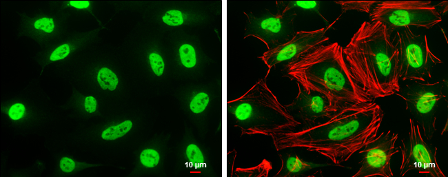
- MAFG antibody detects MAFG protein at nucleus by immunofluorescent analysis.Sample: HeLa cells were fixed in 4% paraformaldehyde at RT for 15 min.Green: MAFG protein stained by MAFG antibody (GTX114541) diluted at 1:500.Red: phalloidin, a cytoskeleton marker, diluted at 1:50.Scale bar = 10 £gm.
Supportive validation
- Submitted by
- GeneTex (provider)
- Main image
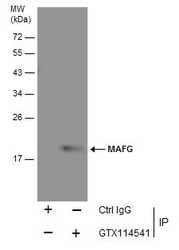
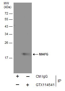
- Experimental details
- Immunoprecipitation of MAFG protein from HeLa whole cell extracts using 5 £gg of MAFG antibody (GTX114541) .Western blot analysis was performed using MAFG antibody (GTX114541).EasyBlot anti-Mouse IgG (GTX221667-01) was used as a secondary reagent.
Supportive validation
- Submitted by
- GeneTex (provider)
- Main image
- Experimental details
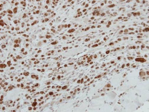
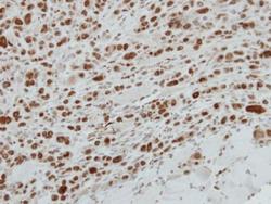
- Immunohistochemical analysis of paraffin-embedded PC9 xenograft, using MAFG(GTX114541) antibody at 1:500 dilution.