Explore
 Validate
 Learn
Learn Western blot
Antibody data
- Antibody Data
- Antigen structure
- References [0]
- Comments [0]
- Validations
- Western blot [2]
- Immunocytochemistry [1]
Submit
Validation data
Reference
Comment
Report error
- Product number
- 701988 - Provider product page

- Provider
- Invitrogen Antibodies
- Product name
- GLT-1 Recombinant Rabbit Monoclonal Antibody (9H9L17)
- Antibody type
- Monoclonal
- Antigen
- Synthetic peptide
- Description
- This antibody is predicted to react with Monkey, Rabbit and Sheep
- Antibody clone number
- 9H9L17
- Concentration
- 0.5 mg/mL
No comments: Submit comment
Supportive validation
- Submitted by
- Invitrogen Antibodies (provider)
- Main image

- Experimental details
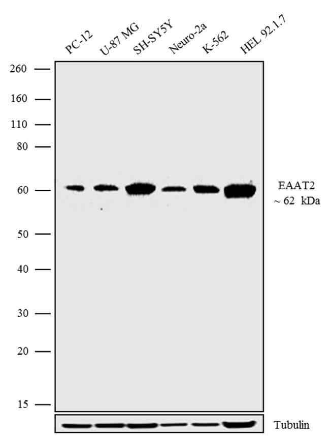
- Western blot analysis was performed on whole cell extracts (30 µg lysate) of PC-12 (Lane 1), U-87 MG (Lane 2), SH-SY-5Y (Lane 3), Neuro-2a (Lane 4), K-562 (Lane 5), and HEL 92.1.7 (Lane 6). The blots were probed with Anti-EAAT2 Recombinant Rabbit Monoclonal Antibody (Product # 701988, 1-2 µg/mL) and detected by chemiluminescence using Goat anti-Rabbit IgG (H+L) Superclonal™ Secondary Antibody, HRP conjugate (Product # A27036, 0.4 µg/mL, 1:2500 dilution). A 62 kDa band corresponding to EAAT2 was observed across cell lines tested. Known quantity of protein samples were electrophoresed using Novex® NuPAGE® 4-12% Bis-Tris gel (Product # NP0321BOX), XCell SureLock™ Electrophoresis System (Product # EI0002) and Novex® Sharp Pre-Stained Protein Standard (Product # LC5800). Resolved proteins were then transferred onto a nitrocellulose membrane with iBlot® Dry Blotting System (Product # IB21001). The membrane was probed with the relevant primary and secondary Antibody following blocking with 5% skimmed milk. Chemiluminescent detection was performed using Pierce™ ECL Western blotting Substrate (Product # 32106).
- Submitted by
- Invitrogen Antibodies (provider)
- Main image

- Experimental details
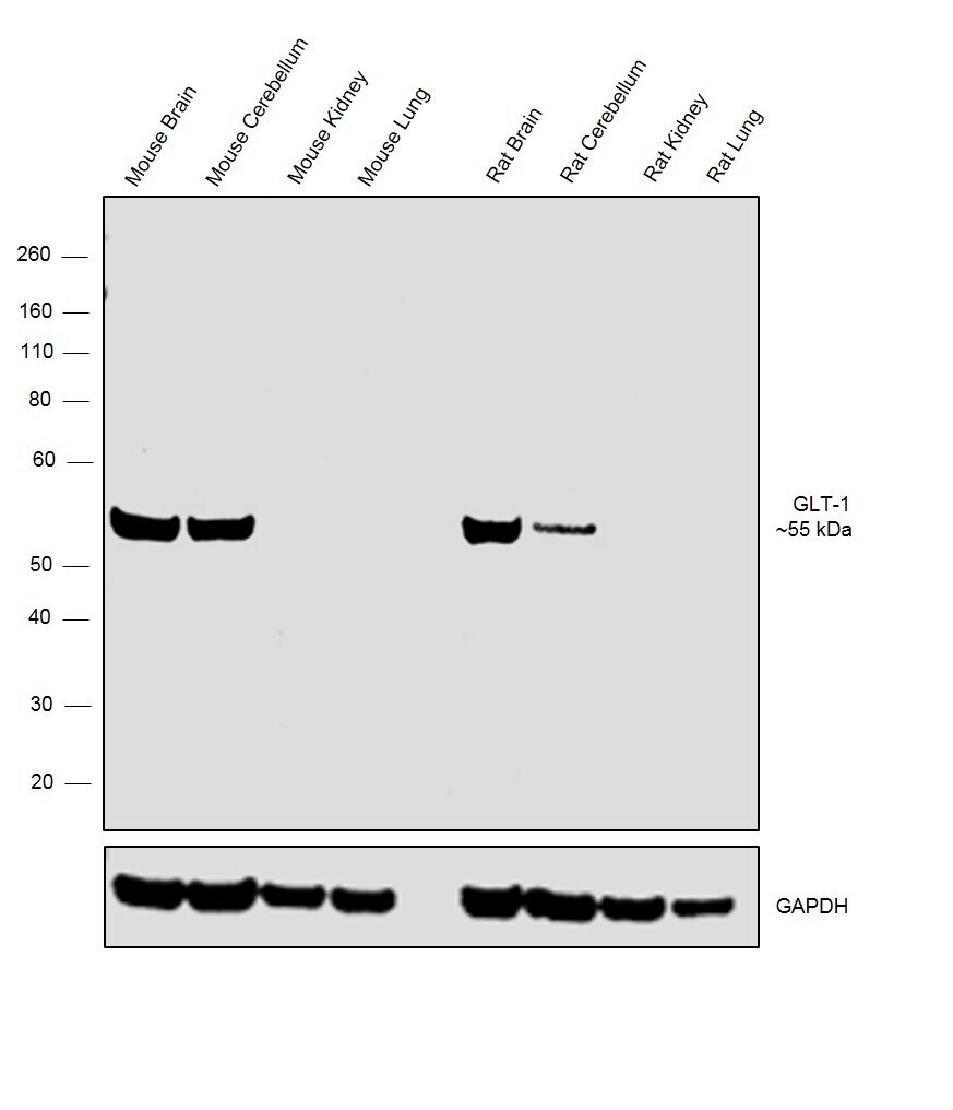
- Western blot was performed using Anti-GLT-1 Monoclonal Antibody, (Product # 701988) and band at ~55 kDa corresponding to GLT-1 was observed in Mouse and Rat Brain and Cerebellum but not in Kidney and Lung which are reported to be negative. Tissue extracts (30 µg lysate) of Mouse Brain (Lane 1), Mouse Cerebellum (Lane 2), Mouse Kidney (Lane 3), Mouse Lung (Lane 4), Rat Brain (Lane 5), Rat Cerebellum (Lane 6), Rat Kidney (Lane 7) and Rat Lung (Lane 8) were electrophoresed using Novex® NuPAGE® 4-12% % Bis-Tris gel (Product # NP0322BOX). Resolved proteins were then transferred onto a nitrocellulose membrane (Product # IB23001) by iBlot® 2 Dry Blotting System (Product # IB21001). The blot was probed with the primary antibody (2µg/ml) and detected by chemiluminescence with Goat anti- Rabbit IgG (H+L) Superclonal™ Recombinant Secondary Antibody, HRP (Product # A27036, 1:4000 dilution) using the iBright FL 1000 (Product # A32752). Chemiluminescent detection was performed using Novex® ECL Chemiluminescent Substrate Reagent Kit (Product # WP20005)..
Supportive validation
- Submitted by
- Invitrogen Antibodies (provider)
- Main image
- Experimental details
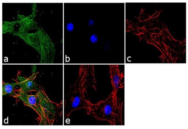
- Immunofluorescence analysis was performed on fixed and permeabilized U-87 MG cells for detection of endogenous EAAT2 using Anti-EAAT2 Recombinant Rabbit Monoclonal Antibody (Product # 701988, 2 µg/mL) and labeled with Goat anti-Rabbit IgG (H+L) Superclonal™ Secondary Antibody, Alexa Fluor® 488 conjugate (Product # A27034, 1:2000). Panel a) shows representative cells that were stained for detection and localization of EAAT2 protein (green), Panel b) is stained for nuclei (blue) using SlowFade® Gold Antifade Mountant with DAPI (Product # S36938). Panel c) represents cytoskeletal F-actin staining using Alexa Fluor® 555 Rhodamine Phalloidin (Product # R415, 1:300). Panel d) is a composite image of Panels a, b and c clearly demonstrating localization of EAAT2 in the membrane. Panel e) represents control cells with no primary antibody to assess background. The images were captured at 60X magnification.