Explore
 Validate
 Learn
Learn Western blot
 Immunocytochemistry
Antibody data
- Antibody Data
- Antigen structure
- References [0]
- Comments [0]
- Validations
- Western blot [5]
- Immunoprecipitation [1]
- Immunohistochemistry [2]
Submit
Validation data
Reference
Comment
Report error
- Product number
- NBP1-33684 - Provider product page

- Provider
- Novus Biologicals
- Proper citation
- Novus Cat#NBP1-33684, RRID:AB_10004096
- Product name
- Rabbit Polyclonal Annexin A1 Antibody
- Antibody type
- Polyclonal
- Description
- Immunogen affinity purified.
- Reactivity
- Human, Mouse, Rat
- Host
- Rabbit
- Isotype
- IgG
- Vial size
- 0.1 ml
- Storage
- Aliquot and store at -20C or -80C. Avoid freeze-thaw cycles.
No comments: Submit comment
Supportive validation
- Submitted by
- Novus Biologicals (provider)
- Main image

- Experimental details
- Western Blot: Annexin A1 Antibody [NBP1-33684] - Sample (30 ug of whole cell lysate) A: NIH-3T3 B: JC 10% SDS PAGE; antibody diluted at 1:10000.

- Submitted by
- Novus Biologicals (provider)
- Main image

- Experimental details
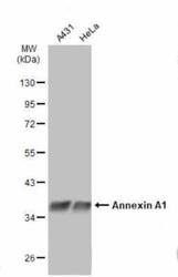
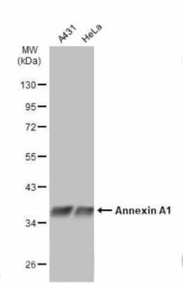
- Western Blot: Annexin A1 Antibody [NBP1-33684] - Sample (30 ug of whole cell lysate) A: A431 B: H1299 C: Hela 10% SDS PAGE; antibody diluted at 1:1000.

- Submitted by
- Novus Biologicals (provider)
- Main image
- Experimental details
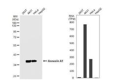
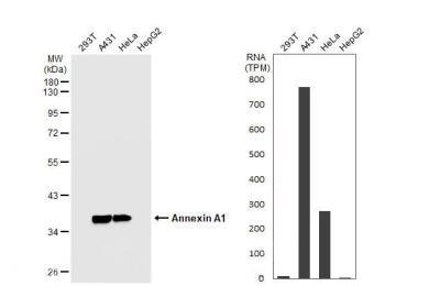
- Western Blot: Annexin A1 Antibody [NBP1-33684] - Various whole cell extracts (30 ug) were separated by 10% SDS-PAGE, and the membrane was blotted with Annexin A1 antibody diluted at 1:10000. HRP-conjugated anti-rabbit IgG antibody was used to detect the primary antibody.
- Submitted by
- Novus Biologicals (provider)
- Main image
- Experimental details
- Western Blot: Annexin A1 Antibody [NBP1-33684] - Various whole cell extracts (30 ug) were separated by 10% SDS-PAGE, and the membranes were blotted with Annexin A1 antibody diluted at 1:5000. HRP-conjugated anti-rabbit IgG antibody was used to detect the primary antibody.
- Submitted by
- Novus Biologicals (provider)
- Main image

- Experimental details
- Western Blot: Annexin A1 Antibody [NBP1-33684] - Non-transfected (-) and transfected (+) 293T whole cell extracts (30 ug) were separated by 10% SDS-PAGE, and the membrane was blotted with Annexin A1 antibody diluted at 1:10000. HRP-conjugated anti-rabbit IgG antibody was used to detect the primary antibody.

Supportive validation
- Submitted by
- Novus Biologicals (provider)
- Main image

- Experimental details
- Immunoprecipitation: Annexin A1 Antibody [NBP1-33684] - Immunoprecipitation of Annexin A1 protein from HeLa whole cell extracts using 5 ug of Annexin A1 antibody. Western blot analysis was performed using Annexin A1 antibody. EasyBlot anti-Rabbit IgG was used as a secondary reagent.

Supportive validation
- Submitted by
- Novus Biologicals (provider)
- Main image
- Experimental details
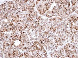
- Immunohistochemistry-Paraffin: Annexin A1 Antibody [NBP1-33684] - Paraffin-embedded N87 Xenograft, using antibody at 1:500 dilution.

- Submitted by
- Novus Biologicals (provider)
- Main image

- Experimental details
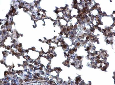
- Immunohistochemistry-Paraffin: Annexin A1 Antibody [NBP1-33684] - Paraffin-embedded mouse lung. Annexin A1 antibody dilution: 1:500.