 Explore
 Validate
 Learn
Learn Western blot
 Flow cytometry
Antibody data
- Antibody Data
- Antigen structure
- References [5]
- Comments [0]
- Validations
- Western blot [1]
- Immunocytochemistry [1]
Submit
Validation data
Reference
Comment
Report error
- Product number
- MAB932-100 - Provider product page

- Provider
- R&D Systems
- Product name
- Human MCAM/CD146 Antibody
- Antibody type
- Monoclonal
- Description
- Protein A or G purified from hybridoma culture supernatant. Detects human MCAM in direct ELISAs and Western blots. In direct ELISAs and Western blots, no cross-reactivity with recombinant human (rh) ALCAM, rhBCAM, rhEpCAM, rhNCAM-L1, recombinant mouse (rm) OCAM, or rmMAdCAM-1 is observed.
- Reactivity
- Human
- Host
- Mouse
- Conjugate
- Unconjugated
- Antigen sequence
AAA20824- Isotype
- IgG
- Antibody clone number
- 128018
- Vial size
- 100 ug
- Storage
- Use a manual defrost freezer and avoid repeated freeze-thaw cycles. 12 months from date of receipt, -20 to -70 °C as supplied. 1 month, 2 to 8 °C under sterile conditions after reconstitution. 6 months, -20 to -70 °C under sterile conditions after reconstitution.
Submitted references Fluoxetine induces direct inhibitory effects on mesenchymal stem cell‑derived osteoprogenitor cells independent of serotonin concentration.
Methods for Systematic Identification of Membrane Proteins for Specific Capture of Cancer-Derived Extracellular Vesicles.
Feasibility and Efficiency of Human Bone Marrow Stromal Cell Culture with Allogeneic Platelet Lysate-Supplementation for Cell Therapy against Stroke.
Choice of xenogenic-free expansion media significantly influences the myogenic differentiation potential of human bone marrow-derived mesenchymal stromal cells.
Label-free detection and molecular profiling of exosomes with a nano-plasmonic sensor.
Koura SM, Salama M, El-Hussiny M, Khalil MEA, Lotfy A, Hassan SA, Gad Elhak SA, Sobh MA
Molecular medicine reports 2019 Apr;19(4):2611-2619
Molecular medicine reports 2019 Apr;19(4):2611-2619
Methods for Systematic Identification of Membrane Proteins for Specific Capture of Cancer-Derived Extracellular Vesicles.
Zaborowski MP, Lee K, Na YJ, Sammarco A, Zhang X, Iwanicki M, Cheah PS, Lin HY, Zinter M, Chou CY, Fulci G, Tannous BA, Lai CP, Birrer MJ, Weissleder R, Lee H, Breakefield XO
Cell reports 2019 Apr 2;27(1):255-268.e6
Cell reports 2019 Apr 2;27(1):255-268.e6
Feasibility and Efficiency of Human Bone Marrow Stromal Cell Culture with Allogeneic Platelet Lysate-Supplementation for Cell Therapy against Stroke.
Tan C, Shichinohe H, Wang Z, Hamauchi S, Abumiya T, Nakayama N, Kazumata K, Ito T, Kudo K, Takamoto S, Houkin K
Stem cells international 2016;2016:6104780
Stem cells international 2016;2016:6104780
Choice of xenogenic-free expansion media significantly influences the myogenic differentiation potential of human bone marrow-derived mesenchymal stromal cells.
Brun J, Abruzzese T, Rolauffs B, Aicher WK, Hart ML
Cytotherapy 2016 Mar;18(3):344-59
Cytotherapy 2016 Mar;18(3):344-59
Label-free detection and molecular profiling of exosomes with a nano-plasmonic sensor.
Im H, Shao H, Park YI, Peterson VM, Castro CM, Weissleder R, Lee H
Nature biotechnology 2014 May;32(5):490-5
Nature biotechnology 2014 May;32(5):490-5
No comments: Submit comment
Supportive validation
- Submitted by
- R&D Systems (provider)
- Main image

- Experimental details
- Detection of Human MCAM/CD146 by Western Blot. Western blot shows lysate of HeLa human cervical epithelial carcinoma cell line. PVDF membrane was probed with 1 µg/mL of Mouse Anti-Human MCAM/CD146 Monoclonal Antibody (Catalog # MAB932) followed by HRP-conjugated Anti-Mouse IgG Secondary Antibody (Catalog # HAF018). A specific band was detected for MCAM/CD146 at approximately 140 kDa (as indicated). This experiment was conducted under reducing conditions and using Immunoblot Buffer Group 1.

Supportive validation
- Submitted by
- R&D Systems (provider)
- Main image
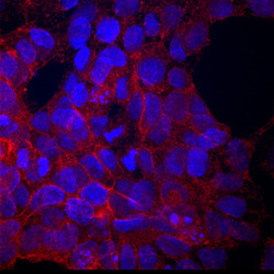
- Experimental details
- MCAM/CD146 in BG01V Human Embryonic Stem Cells. MCAM/CD146 was detected in immersion fixed BG01V human embryonic stem cells using Mouse Anti-Human MCAM/CD146 Monoclonal Antibody (Catalog # MAB932) at 10 µg/mL for 3 hours at room temperature. Cells were stained using the NorthernLights™ 557-conjugated Anti-Mouse IgG Secondary Antibody (red; Catalog # NL007) and counterstained with DAPI (blue). Specific staining was localized to cell membranes and cytoplasm. View our protocol for Fluorescent ICC Staining of Stem Cells on Coverslips.
