Explore
 Validate
 Learn
Learn Western blot
Antibody data
- Antibody Data
- Antigen structure
- References [0]
- Comments [0]
- Validations
- Western blot [7]
- Immunocytochemistry [2]
- Immunohistochemistry [1]
Submit
Validation data
Reference
Comment
Report error
- Product number
- PA5-21583 - Provider product page

- Provider
- Invitrogen Antibodies
- Product name
- TPI1 Polyclonal Antibody
- Antibody type
- Polyclonal
- Antigen
- Synthetic peptide
- Description
- Recommended positive controls: 293T, A431, HeLa, HepG2, Neuro2A, GL261, C8D30, NIH-3T3, BCL-1, Raw264.7, C2C12, PC-12, Rat2.
- Concentration
- 0.27 mg/mL
No comments: Submit comment
Supportive validation
- Submitted by
- Invitrogen Antibodies (provider)
- Main image
- Experimental details
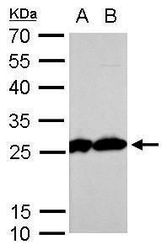
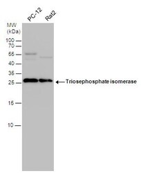
- Western blot analysis of TPI1 using A) 30 µg PC-12 whole cell lysate and B) 30 µg Rat2 whole cell lysate. Samples were loaded onto a 12% SDS-PAGE gel and probed with a TPI1 polyclonal antibody (Product # PA5-21583) at a dilution of 1:1000.

- Submitted by
- Invitrogen Antibodies (provider)
- Main image
- Experimental details
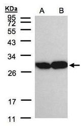
- Western blot analysis of TPI1 using 30 µg of A) A431 and B) H1299 lysate. Samples were loaded onto a 12% SDS-PAGE gel and probed with a TPI1 polyclonal antibody (Product # PA5-21583) at a dilution of 1:2000.
- Submitted by
- Invitrogen Antibodies (provider)
- Main image

- Experimental details
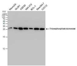
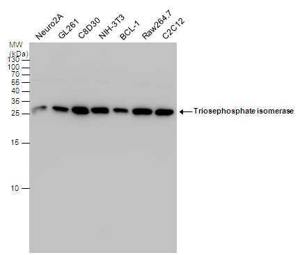
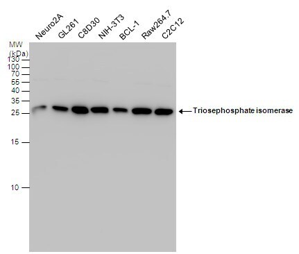
- Western blot analysis of TPI1 using A) 30 µg Neuro2A whole cell lysate (B) 30 µg GL261 whole cell lysate (C) 30 µg C8D30 whole cell lysate (D) 30 µg NIH-3T3 whole cell lysate (E) 30 µg BCL-1 whole cell lysate (F) 30 µg Raw264.7 whole cell lysate and G) 30 µg C2C12 whole cell lysate. Samples were loaded onto a 12% SDS-PAGE gel and probed with a TPI1 polyclonal antibody (Product # PA5-21583) at a dilution of 1:1000.
- Submitted by
- Invitrogen Antibodies (provider)
- Main image
- Experimental details
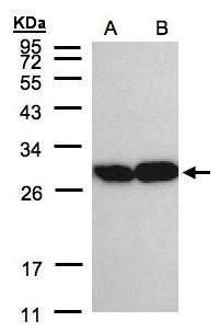
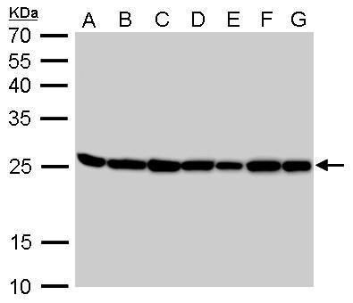
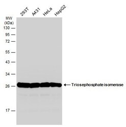
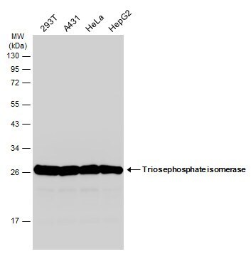
- Western Blot analysis of TPI1 was performed by separating 30 µg of various whole cell extracts by 15% SDS-PAGE. Proteins were transferred to a membrane and probed with a TPI1 Polyclonal Antibody (Product # PA5-21583) at a dilution of 1:1000 and a HRP-conjugated anti-rabbit IgG secondary antibody.
- Submitted by
- Invitrogen Antibodies (provider)
- Main image
- Experimental details
- Western Blot analysis of TPI1 was performed by separating 30 µg of various whole cell extracts by 12% SDS-PAGE. Proteins were transferred to a membrane and probed with a TPI1 Polyclonal Antibody (Product # PA5-21583) at a dilution of 1:1000 and a HRP-conjugated anti-rabbit IgG secondary antibody.
- Submitted by
- Invitrogen Antibodies (provider)
- Main image

- Experimental details
- Western Blot analysis of TPI1 was performed by separating 30 µg of various whole cell extracts by 15% SDS-PAGE. Proteins were transferred to a membrane and probed with a TPI1 Polyclonal Antibody (Product # PA5-21583) at a dilution of 1:1000 and a HRP-conjugated anti-rabbit IgG secondary antibody.
- Submitted by
- Invitrogen Antibodies (provider)
- Main image
- Experimental details
- Western Blot analysis of TPI1 was performed by separating 30 µg of various whole cell extracts by 15% SDS-PAGE. Proteins were transferred to a membrane and probed with a TPI1 Polyclonal Antibody (Product # PA5-21583) at a dilution of 1:1000 and a HRP-conjugated anti-rabbit IgG secondary antibody.

Supportive validation
- Submitted by
- Invitrogen Antibodies (provider)
- Main image
- Experimental details
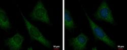
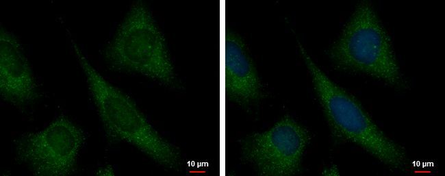
- Immunofluorescent analysis of TPI1 showing staining in the cytoplasm of HeLa cells. HeLa cells were fixed in 4% paraformaldehyde at RT for 15 min and stained using a TPI1 polyclonal antibody (Product # PA5-21583) diluted at 1:500. Blue: Hoechst 33342 staining.
- Submitted by
- Invitrogen Antibodies (provider)
- Main image
- Experimental details
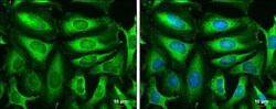
- Immunocytochemistry-Immunofluorescence analysis of TPI1 was performed in HeLa cells fixed in 4% paraformaldehyde at RT for 15 min. Green: TPI1 Polyclonal Antibody (Product # PA5-21583) diluted at 1:500. Blue: Hoechst 33342 staining. Scale bar = 10 µm.

Supportive validation
- Submitted by
- Invitrogen Antibodies (provider)
- Main image
- Experimental details
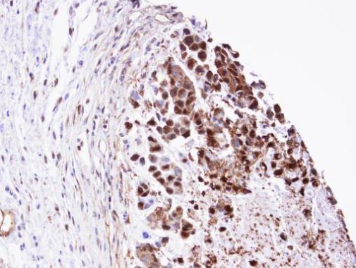
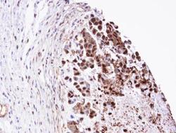
- Immunohistochemical analysis of paraffin-embedded SNU-16 xenograft, using TPI1 (Product # PA5-21583) antibody at 1:100 dilution. Antigen Retrieval: EDTA based buffer, pH 8.0, 15 min.