Explore
 Validate
 Learn
Learn Western blot
Antibody data
- Antibody Data
- Antigen structure
- References [0]
- Comments [0]
- Validations
- Western blot [4]
- Immunocytochemistry [1]
- Immunohistochemistry [2]
Submit
Validation data
Reference
Comment
Report error
- Product number
- PA5-19958 - Provider product page

- Provider
- Invitrogen Antibodies
- Product name
- BNIP3L Polyclonal Antibody
- Antibody type
- Polyclonal
- Antigen
- Synthetic peptide
- Description
- In Western blot, an upper band is detected at ~40kDa, which represents the monomer of Bnip3L. A suggested positive control is K562 cell lysate.
- Concentration
- 1 mg/mL
No comments: Submit comment
Supportive validation
- Submitted by
- Invitrogen Antibodies (provider)
- Main image
- Experimental details
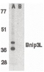
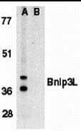
- Western blot analysis of K562 whole cell lysate in the absence (A), or presence (B) of immunogenic peptide using a Bnip3L polyclonal antibody (Product # PA5-19958) at 1 µg/mL.
- Submitted by
- Invitrogen Antibodies (provider)
- Main image

- Experimental details
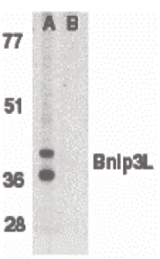
- Western Blot analysis of Bnip3L in K562 whole cell lysate in (A) the absence, or (B) presence of immunogenic peptide with BNIP3L Polyclonal Antibody (Product # PA5-19958) at 1 µg/mL.
- Submitted by
- Invitrogen Antibodies (provider)
- Main image

- Experimental details
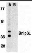
- Western Blot analysis of Bnip3L in K562 whole cell lysate in (A) the absence, or (B) presence of immunogenic peptide with BNIP3L Polyclonal Antibody (Product # PA5-19958) at 1 µg/mL.
- Submitted by
- Invitrogen Antibodies (provider)
- Main image
- Experimental details
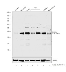
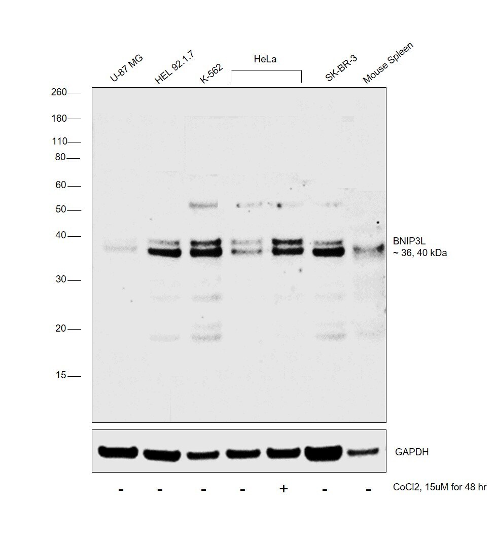
- Western blot was performed using Anti-BNIP3L Polyclonal Antibody (Product # PA5-19958) and 36, 40 kDa bands corresponding to BNIP3L were observed across all the cell lines and tissues tested and was observed to increase upon Cobalt chloride treatment in HeLa. Whole cell extracts (30 µg lysate) of U-87 MG (Lane 1), HEL 92.1.7 (Lane 2), K-562 (Lane 3), HeLa (Lane 4), HeLa treated with Cobalt Chloride (15uM for 48hr) (Lane 5), SK-BR-3 (Lane 6) and tissue extract of Mouse Spleen (Lane 7) were electrophoresed using Novex® NuPAGE® 4-12% Bis-Tris Protein Gel (Product # NP0322BOX). Resolved proteins were then transferred onto a nitrocellulose membrane (Product # IB23001) by iBlot® 2 Dry Blotting System (Product # IB21001). The blot was probed with the primary antibody (1ug/ml) and detected by chemiluminescence with Goat anti-Rabbit IgG (H+L), Superclonal™ Recombinant Secondary Antibody, HRP (Product # A27036, 1:4000 dilution) using the iBright FL 1000 (Product # A32752). Chemiluminescent detection was performed using Novex® ECL Chemiluminescent Substrate Reagent Kit (Product # WP20005).
Supportive validation
- Submitted by
- Invitrogen Antibodies (provider)
- Main image
- Experimental details
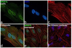
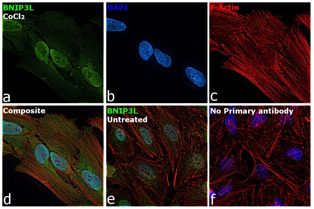
- Immunofluorescence analysis of BNIP3L was performed using 70% confluent log phase HeLa cells treated with Cobalt chloride. The cells were fixed with 4% paraformaldehyde for 10 minutes, permeabilized with 0.1% Triton™ X-100 for 15 minutes, and blocked with 2% BSA for 1 hour at room temperature. The cells were labeled with PHD1 Recombinant Rabbit Monoclonal Antibody (3G4) (Product # PA5-19958) at 5 µg/mL dilution in 0.1% BSA, incubated at 4 degree Celsius overnight and then labeled with Goat anti-Rabbit IgG (H+L) Superclonal™ Recombinant Secondary Antibody, Alexa Fluor® 488 conjugate (Product # A27034) at a dilution of 1:2000 for 45 minutes at room temperature (Panel a: green). Nuclei (Panel b: blue) were stained with ProLong™ Diamond Antifade Mountant with DAPI (Product # P36962). F-actin (Panel c: red) was stained with Rhodamine Phalloidin (Product # R415). Panel d represents the merged image showing increase in Nuclear localization. Panel e represents untreated cells with Nuclear localization. Panel f represents control cells with no primary antibody to assess background. The images were captured at 60X magnification.
Supportive validation
- Submitted by
- Invitrogen Antibodies (provider)
- Main image
- Experimental details
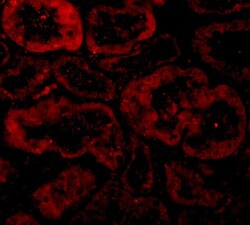
- Immunofluorescence of Bnip3L in Human Kidney tissue with BNIP3L Polyclonal Antibody (Product # PA5-19958) at 10 µg/mL.
- Submitted by
- Invitrogen Antibodies (provider)
- Main image

- Experimental details
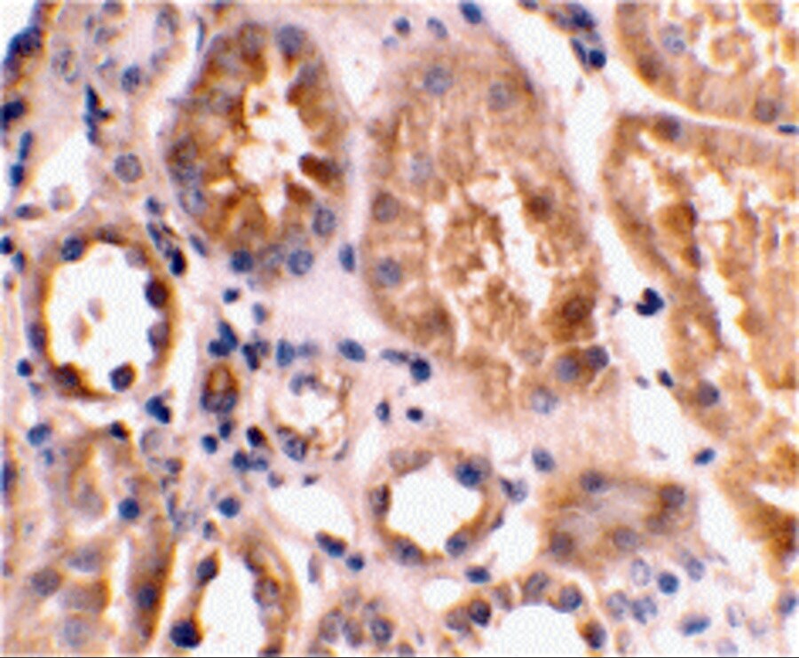
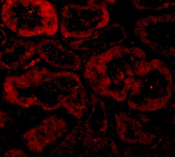
- Immunohistochemical staining of human kidney tissue using BNIP3L Polyclonal Antibody (Product # PA5-19958) at 2 µg/mL.