 Explore
 Validate
 Learn
Learn ELISA
ELISAAntibody data
- Antibody Data
- Antigen structure
- References [1]
- Comments [0]
- Validations
- ELISA [1]
- Immunocytochemistry [1]
- Immunohistochemistry [1]
- Proximity ligation assay [1]
Submit
Validation data
Reference
Comment
Report error
- Product number
- H00000821-M08 - Provider product page

- Provider
- Abnova Corporation
- Proper citation
- Abnova Corporation Cat#H00000821-M08, RRID:AB_1137121
- Product name
- CANX monoclonal antibody (M08), clone 1D4
- Antibody type
- Monoclonal
- Description
- Mouse monoclonal antibody raised against a partial recombinant CANX.
- Antigen sequence
SGKKQTSGMEYKKTDAPQPDVKEEEEEKEEEKDKG
DEEEEGEEKLEEKQKSDAEEDGGTVSQEEEDRKPK
AEEDEILNRSPRNRKPRRE- Isotype
- IgG
- Antibody clone number
- 1D4
- Storage
- Store at -20°C or lower. Aliquot to avoid repeated freezing and thawing.
Submitted references The SARS coronavirus E protein interacts with PALS1 and alters tight junction formation and epithelial morphogenesis.
Teoh KT, Siu YL, Chan WL, Schlüter MA, Liu CJ, Peiris JS, Bruzzone R, Margolis B, Nal B
Molecular biology of the cell 2010 Nov 15;21(22):3838-52
Molecular biology of the cell 2010 Nov 15;21(22):3838-52
No comments: Submit comment
Supportive validation
- Submitted by
- Abnova Corporation (provider)
- Main image
- Experimental details
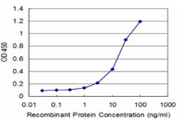
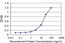
- Detection limit for recombinant GST tagged CANX is approximately 1ng/ml as a capture antibody.
- Validation comment
- Sandwich ELISA (Recombinant protein)
- Protocol
- Protocol
Supportive validation
- Submitted by
- Abnova Corporation (provider)
- Main image

- Experimental details
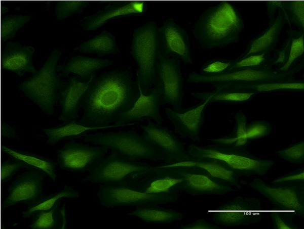
- Immunofluorescence of monoclonal antibody to CANX on HeLa cell . [antibody concentration 10 ug/ml]
- Validation comment
- Immunofluorescence
- Protocol
- Protocol
Supportive validation
- Submitted by
- Abnova Corporation (provider)
- Main image

- Experimental details
- Immunoperoxidase of monoclonal antibody to CANX on formalin-fixed paraffin-embedded human placenta. [antibody concentration 1.5 ug/ml]
- Validation comment
- Immunohistochemistry (Formalin/PFA-fixed paraffin-embedded sections)
- Protocol
- Protocol

Supportive validation
- Submitted by
- Abnova Corporation (provider)
- Main image
- Experimental details
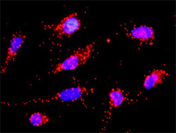
- Proximity Ligation Analysis of protein-protein interactions between CD3D and CANX. HeLa cells were stained with anti-CD3D rabbit purified polyclonal 1:1200 and anti-CANX mouse monoclonal antibody 1:50. Each red dot represents the detection of protein-protein interaction complex, and nuclei were counterstained with DAPI (blue).
- Validation comment
- In situ Proximity Ligation Assay (Cell)
