Explore
 Validate
 Learn
Learn Immunocytochemistry
Antibody data
- Antibody Data
- Antigen structure
- References [0]
- Comments [0]
- Validations
- Immunocytochemistry [2]
Submit
Validation data
Reference
Comment
Report error
- Product number
- 703456 - Provider product page

- Provider
- Invitrogen Antibodies
- Product name
- GRSF1 Recombinant Rabbit Monoclonal Antibody (11H24L16)
- Antibody type
- Monoclonal
- Antigen
- Other
- Description
- This antibody is predicted to react with Monkey, Bovine, Pig, Mouse.
- Antibody clone number
- 11H24L16
- Concentration
- 0.5 mg/mL
No comments: Submit comment
Supportive validation
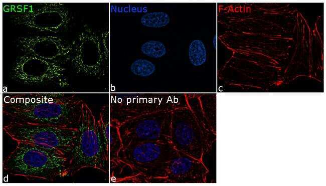
- Submitted by
- Invitrogen Antibodies (provider)
- Main image
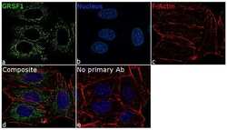
- Experimental details
- For immunofluorescence analysis, MCF7 cells were fixed and permeabilized for detection of endogenous GRSF1 using Anti-GRSF1 Recombinant Rabbit Monoclonal Antibody (Product # 703456, 1:100 dilution) and labeled with Goat anti-Rabbit IgG (H+L) Superclonal™ Secondary Antibody, Alexa Fluor® 488 conjugate (Product # A27034, 1:2000). Panel a) shows representative cells that were stained for detection and localization of GRSF1 protein (green), Panel b) is stained for nuclei (blue) using ProLong™ Diamond Antifade Mountant with DAPI (Product # P36962). Panel c) represents cytoskeletal F-actin staining using Rhodamine Phalloidin (Product # R415, 1:300). Panel d) is a composite image of Panels a, b and c clearly demonstrating mitochondrial localization of GRSF1. Panel e) represents control cells with no primary antibody to assess background. The images were captured at 60X magnification.
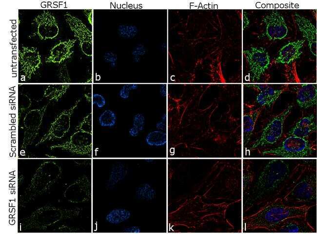
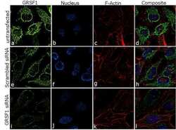
- Submitted by
- Invitrogen Antibodies (provider)
- Main image
- Experimental details
- Knockdown of GRSF1 was achieved by transfecting HeLa cells with specific siRNA (Silencer® select Product # s6234, s6233). Immunofluorescence analysis was performed on HeLa cells (untransfected, panel a-d), transfected with GRSF1 specific siRNA (panel i-l) or non-specific scrambled siRNA (panels e-h). Cells were fixed, permeabilized, and labeled with Anti-GRSF1 Recombinant Rabbit Monoclonal Antibody (Product # 703456, 1:100 dilution), followed by Goat anti-Rabbit IgG (H+L) Superclonal™ Secondary Antibody, Alexa Fluor® 488 conjugate (Product # A27034, 1:2000). Nuclei (blue) were stained using ProLong™ Diamond Antifade Mountant with DAPI (Product # P36962), and Rhodamine Phalloidin (Product # R415, 1:300) was used for cytoskeletal F-actin (red) staining. Significant reduction of signal was observed upon siRNA mediated knockdown (panel i-l) confirming specificity of the antibody to GRSF1 (green). The images were captured at 60X magnification.