 Explore
 Validate
 Learn
Learn Western blot
Antibody data
- Antibody Data
- Antigen structure
- References [0]
- Comments [0]
- Validations
- Western blot [3]
- Immunocytochemistry [1]
- Immunohistochemistry [6]
Submit
Validation data
Reference
Comment
Report error
- Product number
- PA5-27198 - Provider product page

- Provider
- Invitrogen Antibodies
- Product name
- ABAT Polyclonal Antibody
- Antibody type
- Polyclonal
- Antigen
- Recombinant protein fragment
- Description
- Recommended positive controls: SK-N-SH, mouse brain, rat brain.
- Concentration
- 1 mg/mL
No comments: Submit comment
Supportive validation
- Submitted by
- Invitrogen Antibodies (provider)
- Main image

- Experimental details
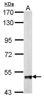
- Western blot analysis of ABAT using 20 µg of mouse brain lysate. Samples were loaded onto a 7.5% SDS-PAGE gel and probed with an ABAT polyclonal antibody (Product # PA5-27198) at a dilution of 1:5000.
- Submitted by
- Invitrogen Antibodies (provider)
- Main image
- Experimental details
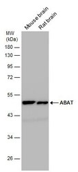
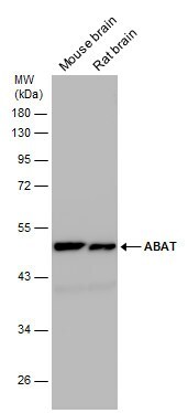
- Western blot analysis of ABAT was performed by separating 50 µg of various tissue extracts by 10% SDS-PAGE. Proteins were transferred to a membrane and probed with a ABAT Polyclonal Antibody (Product # PA5-27198) at a dilution of 1:10000. The HRP-conjugated anti-rabbit IgG antibody was used to detect the primary antibody.
- Submitted by
- Invitrogen Antibodies (provider)
- Main image
- Experimental details
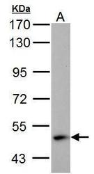
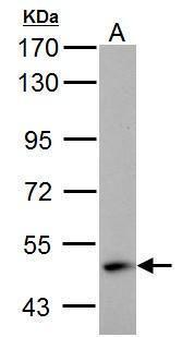
- ABAT Polyclonal Antibody detects ABAT protein by western blot analysis. A. 30 µg SK-N-SH whole cell lysate/extract.7.5% SDS-PAGE. ABAT Polyclonal Antibody (Product # PA5-27198) dilution: 1:1,000. The HRP-conjugated anti-rabbit IgG antibody was used to detect the primary antibody.
Supportive validation
- Submitted by
- Invitrogen Antibodies (provider)
- Main image
- Experimental details
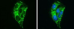
- Immunocytochemistry-Immunofluorescence analysis of ABAT was performed in HepG2 cells fixed in ice-cold MeOH for 5 min. Green: ABAT Polyclonal Antibody (Product # PA5-27198) diluted at 1:500. Blue: Hoechst 33342 staining.

Supportive validation
- Submitted by
- Invitrogen Antibodies (provider)
- Main image
- Experimental details
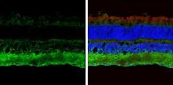
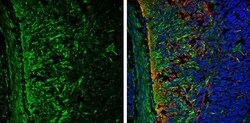
- Immunohistochemistry (Frozen) analysis of ABAT was performed in frozen sectioned adult mouse retina tissue using ABAT Polyclonal Antibody (Product # PA5-27198) at a dilution of 1:250 (Green). Red: beta Tubulin 3/ TUJ1, stained by beta Tubulin 3/ TUJ1 antibody diluted at 1:250. Blue: Fluoroshield with DAPI.
- Submitted by
- Invitrogen Antibodies (provider)
- Main image
- Experimental details
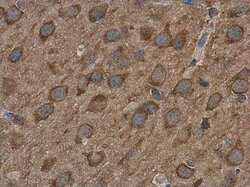
- Immunohistochemistry (Paraffin) analysis of ABAT was performed in paraffin-embedded rat brain tissue using ABAT Polyclonal Antibody (Product # PA5-27198) at a dilution of 1:500.
- Submitted by
- Invitrogen Antibodies (provider)
- Main image
- Experimental details
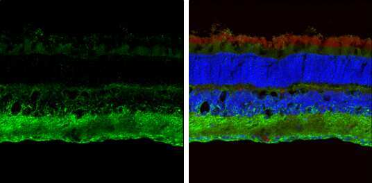
- Immunohistochemistry (Frozen) analysis of ABAT was performed in frozen sectioned E13.5 Rat brain tissue using ABAT Polyclonal Antibody (Product # PA5-27198) at a dilution of 1:250 (Green). Red: beta Tubulin 3/ TUJ1, a mature neuron marker, stained by beta Tubulin 3/ TUJ1 antibody diluted at 1:500. Blue: Fluoroshield with DAPI.

- Submitted by
- Invitrogen Antibodies (provider)
- Main image

- Experimental details
- Immunohistochemistry (Frozen) analysis of ABAT was performed in frozen sectioned adult mouse retina tissue using ABAT Polyclonal Antibody (Product # PA5-27198) at a dilution of 1:250 (Green). Red: beta Tubulin 3/ TUJ1, stained by beta Tubulin 3/ TUJ1 antibody diluted at 1:250. Blue: Fluoroshield with DAPI.

- Submitted by
- Invitrogen Antibodies (provider)
- Main image
- Experimental details
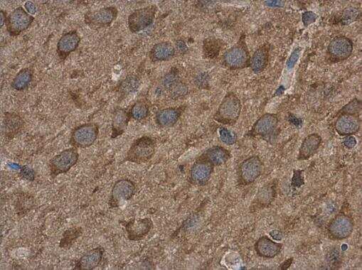
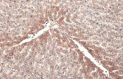
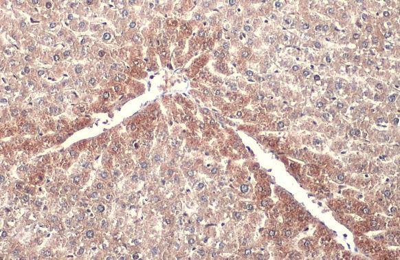
- ABAT Polyclonal Antibody detects ABAT protein at mitochondria by immunohistochemical analysis. Sample: Paraffin-embedded mouse liver. ABAT stained by ABAT Polyclonal Antibody (Product # PA5-27198) diluted at 1:500. Antigen Retrieval: Citrate buffer, pH 6.0, 15 min.
- Submitted by
- Invitrogen Antibodies (provider)
- Main image
- Experimental details
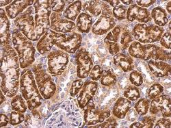
- ABAT Polyclonal Antibody detects ABAT protein at mitochondria on mouse kidney by immunohistochemical analysis. Sample: Paraffin-embedded mouse kidney. ABAT Polyclonal Antibody (Product # PA5-27198) dilution: 1:500. Antigen Retrieval: EDTA based buffer, pH 8.0, 15 min.
