Explore
 Validate
 Learn
Learn Western blot
Antibody data
- Antibody Data
- Antigen structure
- References [2]
- Comments [0]
- Validations
- Western blot [1]
- Immunohistochemistry [1]
- Other assay [3]
Submit
Validation data
Reference
Comment
Report error
- Product number
- PA5-31042 - Provider product page

- Provider
- Invitrogen Antibodies
- Product name
- PCDH10 Polyclonal Antibody
- Antibody type
- Polyclonal
- Antigen
- Recombinant protein fragment
- Description
- Recommended positive controls: NT2D1, PC-3, U87-MG, SK-N-SH.
- Concentration
- 1 mg/mL
Submitted references Promoter methylation of PCDH10 by HOTAIR regulates the progression of gastrointestinal stromal tumors.
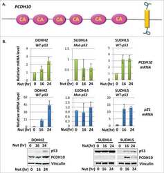
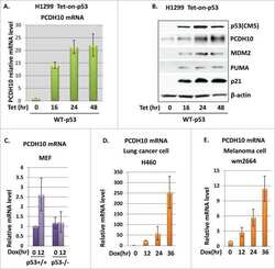
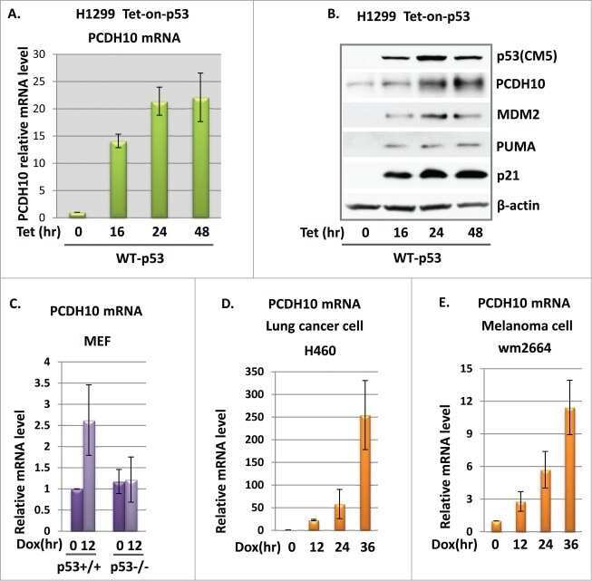
PCDH10, a novel p53 transcriptional target in regulating cell migration.
Lee NK, Lee JH, Kim WK, Yun S, Youn YH, Park CH, Choi YY, Kim H, Lee SK
Oncotarget 2016 Nov 15;7(46):75307-75318
Oncotarget 2016 Nov 15;7(46):75307-75318
PCDH10, a novel p53 transcriptional target in regulating cell migration.
Shi D, Murty VV, Gu W
Cell cycle (Georgetown, Tex.) 2015;14(6):857-66
Cell cycle (Georgetown, Tex.) 2015;14(6):857-66
No comments: Submit comment
Supportive validation
- Submitted by
- Invitrogen Antibodies (provider)
- Main image
- Experimental details
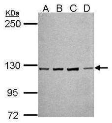
- Western Blot using PCDH10 Polyclonal Antibody (Product # PA5-31042). Sample (30 µg of whole cell lysate). Lane A: NT2D1. Lane B: PC-3. Lane C: U87-MG. Lane D: SK-N-SH . 5% SDS PAGE. PCDH10 Polyclonal Antibody (Product # PA5-31042) diluted at 1:500.

Supportive validation
- Submitted by
- Invitrogen Antibodies (provider)
- Main image

- Experimental details
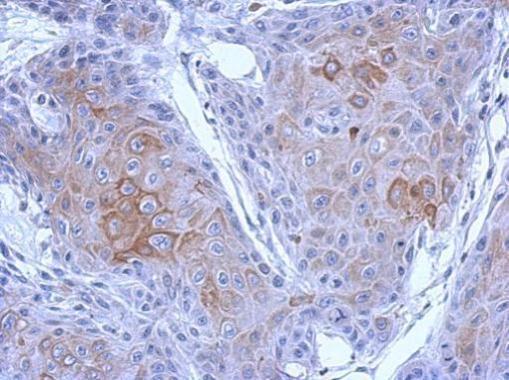
- Immunohistochemical analysis of paraffin-embedded Cal27 xenograft, using PCDH10 (Product # PA5-31042) antibody at 1:500 dilution. Antigen Retrieval: EDTA based buffer, pH 8.0, 15 min.
Supportive validation
- Submitted by
- Invitrogen Antibodies (provider)
- Main image
- Experimental details
- NULL

- Submitted by
- Invitrogen Antibodies (provider)
- Main image
- Experimental details
- NULL
- Submitted by
- Invitrogen Antibodies (provider)
- Main image
- Experimental details
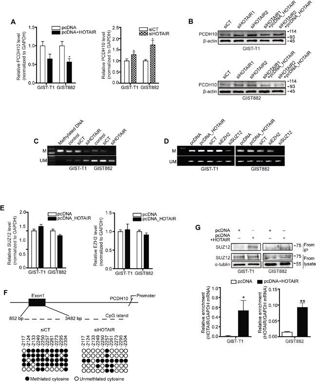
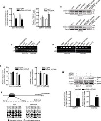
- Figure 3 HOTAIR controls the methylation of PCDH10 PCDH10 expression was measured in GIST-T1 and GIST882 cell lines after treatment with pcDNA-HOTAIR ( A. ; left panel), and in GIST882 cells after treatment with siHOTAIRs (A, right panel). The data shown are representative of three independent experiments. The bars represent relative PCDH10 mRNA levels. B. PCDH10 protein level was determined via Western blot in GIST-T1 and GIST 882 cells. C. The methylation of PCDH10 after treatment with siHOTAIRs was measured via MS-PCR. D. The methylation of PCDH10 was measured after treatment with siEZH2 or siSUZ12. M, methylated DNA; UM, unmethylated DNA. E. mRNA of SUZ12 and EZH2 was measured via qRT-PCR after transfection of pcDNA-HOTAIR. NS: not significant. F. DNA from GIST-T1 treated with siCT or siHOTAIRs was treated with bisulfite, and the promoter regions in axon 1 were amplified by PCR and cloned. Each circle represents a CpG and filled ovals indicate methylation. Open ovals indicate un-methylation. G. The interaction between SUZ12 and HOTAIR was confirmed via RIP analysis. The bars present relative enrichment. Data are expressed as mean +- SEM. The asterisk denotes a statistically significant difference compared to pcDNA or scrambled control (* P